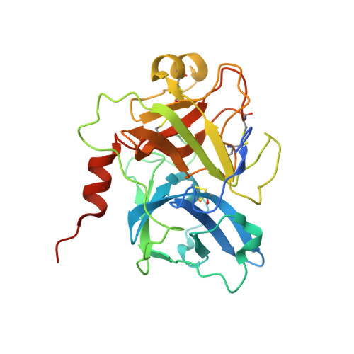
Structures of full-length plasma kallikrein bound to highly specific inhibitors describe a new mode of targeted inhibition.
Partridge, J.R., Choy, R.M., Silva-Garcia, A., Yu, C., Li, Z., Sham, H., Metcalf, B.(2019) J Struct Biol 206: 170-182
- PubMed: 30876891
- DOI: https://doi.org/10.1016/j.jsb.2019.03.001
- Primary Citation of Related Structures:
6BFP, 6O1G, 6O1S - PubMed Abstract:
Plasma kallikrein (pKal) is a serine protease responsible for cleaving high-molecular-weight kininogen to produce the pro-inflammatory peptide, bradykinin. Unregulated pKal activity can lead to hereditary angioedema (HAE) following excess bradykinin release. HAE attacks can lead to a compromised airway that can be life threatening. As there are limited agents for prophylaxis of HAE attacks, there is a high unmet need for a therapeutic agent for regulating pKal with a high degree of specificity. Here we present crystal structures of both full-length and the protease domain of pKal, bound to two very distinct classes of small-molecule inhibitors: compound 1, and BCX4161. Both inhibitors demonstrate low nM inhibitory potency for pKal and varying specificity for related serine proteases. Compound 1 utilizes a surprising mode of interaction and upon binding results in a rearrangement of the binding pocket. Co-crystal structures of pKal describes why this class of small-molecule inhibitor is potent. Lack of conservation in surrounding residues explains the ∼10,000-fold specificity over structurally similar proteases, as shown by in vitro protease inhibition data. Structural information, combined with biochemical and enzymatic analyses, provides a novel scaffold for the design of targeted oral small molecule inhibitors of pKal for treatment of HAE and other diseases resulting from unregulated plasma kallikrein activity.
- Global Blood Therapeutics, South San Francisco, CA 94080, United States. Electronic address: jpartridge@gbt.com.
Organizational Affiliation:


