6OER

Cryo-EM structure of mouse RAG1/2 NFC complex (DNA2)


Experimental Data Snapshot

  • Method: ELECTRON MICROSCOPY
  • Resolution: 3.29 Å
  • Aggregation State: PARTICLE 
  • Reconstruction Method: SINGLE PARTICLE 

wwPDB Validation   3D Report Full Report


This is version 1.3 of the entry. See complete history


Literature

Cutting antiparallel DNA strands in a single active site.

Chen, X.Cui, Y.Best, R.B.Wang, H.Zhou, Z.H.Yang, W.Gellert, M.

(2020) Nat Struct Mol Biol 27: 119-126

  • DOI: https://doi.org/10.1038/s41594-019-0363-2
  • Primary Citation of Related Structures:  
    6OEM, 6OEN, 6OEO, 6OEP, 6OEQ, 6OER, 6V0V

  • PubMed Abstract: 

    A single enzyme active site that catalyzes multiple reactions is a well-established biochemical theme, but how one nuclease site cleaves both DNA strands of a double helix has not been well understood. In analyzing site-specific DNA cleavage by the mammalian RAG1-RAG2 recombinase, which initiates V(D)J recombination, we find that the active site is reconfigured for the two consecutive reactions and the DNA double helix adopts drastically different structures. For initial nicking of the DNA, a locally unwound and unpaired DNA duplex forms a zipper via alternating interstrand base stacking, rather than melting as generally thought. The second strand cleavage and formation of a hairpin-DNA product requires a global scissor-like movement of protein and DNA, delivering the scissile phosphate into the rearranged active site.


  • Organizational Affiliation

    Laboratory of Molecular Biology, NIDDK, National Institutes of Health, Bethesda, MD, USA.


Macromolecules

Find similar proteins by:  (by identity cutoff)  |  3D Structure
Entity ID: 1
MoleculeChains Sequence LengthOrganismDetailsImage
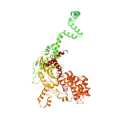
V(D)J recombination-activating protein 1
A, C
1,040Mus musculusMutation(s): 1 
Gene Names: Rag1
EC: 3.1 (PDB Primary Data), 2.3.2.27 (PDB Primary Data)
UniProt
Find proteins for P15919 (Mus musculus)
Explore P15919 
Go to UniProtKB:  P15919
Entity Groups  
Sequence Clusters30% Identity50% Identity70% Identity90% Identity95% Identity100% Identity
UniProt GroupP15919
Sequence Annotations
Expand
  • Reference Sequence
Find similar proteins by:  (by identity cutoff)  |  3D Structure
Entity ID: 2
MoleculeChains Sequence LengthOrganismDetailsImage
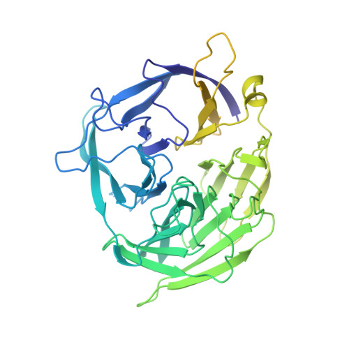
V(D)J recombination-activating protein 2
B, D
527Mus musculusMutation(s): 0 
Gene Names: Rag2Rag-2
UniProt
Find proteins for P21784 (Mus musculus)
Explore P21784 
Go to UniProtKB:  P21784
Entity Groups  
Sequence Clusters30% Identity50% Identity70% Identity90% Identity95% Identity100% Identity
UniProt GroupP21784
Sequence Annotations
Expand
  • Reference Sequence
Find similar proteins by:  (by identity cutoff)  |  3D Structure
Entity ID: 7
MoleculeChains Sequence LengthOrganismDetailsImage
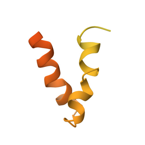
High mobility group protein B1I [auth H]163Equus caballusMutation(s): 0 
UniProt
Find proteins for Q08IE6 (Equus caballus)
Explore Q08IE6 
Go to UniProtKB:  Q08IE6
Entity Groups  
Sequence Clusters30% Identity50% Identity70% Identity90% Identity95% Identity100% Identity
UniProt GroupQ08IE6
Sequence Annotations
Expand
  • Reference Sequence
Find similar nucleic acids by:  (by identity cutoff)  |  3D Structure
Entity ID: 3
MoleculeChains LengthOrganismImage
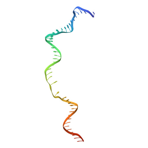
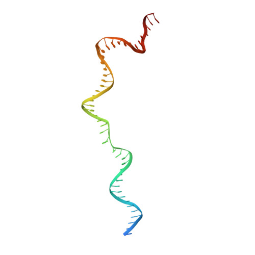
DNA (46-MER)E [auth F]50Escherichia coli K-12
Sequence Annotations
Expand
  • Reference Sequence
Find similar nucleic acids by:  (by identity cutoff)  |  3D Structure
Entity ID: 4
MoleculeChains LengthOrganismImage
DNA (46-MER)F [auth I]50Escherichia coli K-12
Sequence Annotations
Expand
  • Reference Sequence
Find similar nucleic acids by:  (by identity cutoff)  |  3D Structure
Entity ID: 5
MoleculeChains LengthOrganismImage
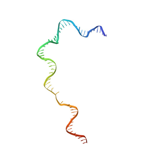
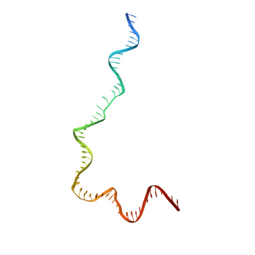
DNA (57-MER)61Escherichia coli K-12
Sequence Annotations
Expand
  • Reference Sequence
Find similar nucleic acids by:  (by identity cutoff)  |  3D Structure
Entity ID: 6
MoleculeChains LengthOrganismImage
DNA (57-MER)H [auth J]61Escherichia coli K-12
Sequence Annotations
Expand
  • Reference Sequence
Experimental Data & Validation

Experimental Data

  • Method: ELECTRON MICROSCOPY
  • Resolution: 3.29 Å
  • Aggregation State: PARTICLE 
  • Reconstruction Method: SINGLE PARTICLE 

Structure Validation

View Full Validation Report


Entry History & Funding Information

Deposition Data


Funding OrganizationLocationGrant Number
National Institutes of Health/National Institute of Diabetes and Digestive and Kidney Disease (NIH/NIDDK)United StatesDK036167

Revision History  (Full details and data files)

  • Version 1.0: 2020-01-29
    Type: Initial release
  • Version 1.1: 2020-02-19
    Changes: Database references
  • Version 1.2: 2020-02-26
    Changes: Database references
  • Version 1.3: 2024-03-20
    Changes: Data collection, Database references, Derived calculations