6OG4

plasminogen binding group A streptococcal M protein


Experimental Data Snapshot

  • Method: X-RAY DIFFRACTION
  • Resolution: 1.70 Å
  • R-Value Free: 0.220 
  • R-Value Work: 0.203 
  • R-Value Observed: 0.204 

wwPDB Validation   3D Report Full Report

Currently 6OG4 does not have a validation slider image.


This is version 1.3 of the entry. See complete history


Literature

Structure and Function Characterization of the a1a2 Motifs of Streptococcus pyogenes M Protein in Human Plasminogen Binding.

Quek, A.J.H.Mazzitelli, B.A.Wu, G.Leung, E.W.W.Caradoc-Davies, T.T.Lloyd, G.J.Jeevarajah, D.Conroy, P.J.Sanderson-Smith, M.Yuan, Y.Ayinuola, Y.A.Castellino, F.J.Whisstock, J.C.Law, R.H.P.

(2019) J Mol Biol 431: 3804-3813

  • DOI: https://doi.org/10.1016/j.jmb.2019.07.003
  • Primary Citation of Related Structures:  
    6OG4

  • PubMed Abstract: 

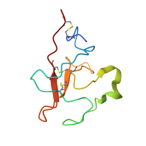
    Plasminogen (Plg)-binding M protein (PAM) is a group A streptococcal cell surface receptor that is crucial for bacterial virulence. Previous studies revealed that, by binding to the kringle 2 (KR2) domain of host Plg, the pathogen attains a proteolytic microenvironment on the cell surface that facilitates its dissemination from the primary infection site. Each of the PAM molecules in their dimeric assembly consists of two Plg binding motifs (called the a1 and a2 repeats). To date, the molecular interactions between the a1 repeat and KR2 have been structurally characterized, whereas the role of the a2 repeat is less well defined. Here, we report the 1.7-Å x-ray crystal structure of KR2 in complex with a monomeric PAM peptide that contains both the a1 and a2 motifs. The structure reveals how the PAM peptide forms key interactions simultaneously with two KR2 via the high-affinity lysine isosteres within the a1a2 motifs. Further studies, through combined mutagenesis and functional characterization, show that a2 is a stronger KR2 binder than a1, suggesting that these two motifs may play discrete roles in mediating the final PAM-Plg assembly.


  • Organizational Affiliation

    ARC Centre of Excellence in Advanced Molecular Imaging, Department of Biochemistry and Molecular Biology, Monash Biomedicine Discovery Institute, Monash University, Clayton, Victoria 3800, Australia.


Macromolecules
Find similar proteins by:  (by identity cutoff)  |  3D Structure
Entity ID: 1
MoleculeChains Sequence LengthOrganismDetailsImage
Plasminogen
A, B
87Homo sapiensMutation(s): 1 
Gene Names: PLG
EC: 3.4.21.7
UniProt & NIH Common Fund Data Resources
Find proteins for P00747 (Homo sapiens)
Explore P00747 
Go to UniProtKB:  P00747
PHAROS:  P00747
GTEx:  ENSG00000122194 
Entity Groups  
Sequence Clusters30% Identity50% Identity70% Identity90% Identity95% Identity100% Identity
UniProt GroupP00747
Sequence Annotations
Expand
  • Reference Sequence
Find similar proteins by:  (by identity cutoff)  |  3D Structure
Entity ID: 2
MoleculeChains Sequence LengthOrganismDetailsImage
Plasminogen-binding group A streptococcal M-like protein PAM77Streptococcus pyogenesMutation(s): 0 
Gene Names: pamemm
UniProt
Find proteins for P49054 (Streptococcus pyogenes)
Explore P49054 
Go to UniProtKB:  P49054
Entity Groups  
Sequence Clusters30% Identity50% Identity70% Identity90% Identity95% Identity100% Identity
UniProt GroupP49054
Sequence Annotations
Expand
  • Reference Sequence
Experimental Data & Validation

Experimental Data

  • Method: X-RAY DIFFRACTION
  • Resolution: 1.70 Å
  • R-Value Free: 0.220 
  • R-Value Work: 0.203 
  • R-Value Observed: 0.204 
  • Space Group: P 43 21 2
Unit Cell:
Length ( Å )Angle ( ˚ )
a = 52.599α = 90
b = 52.599β = 90
c = 198.25γ = 90
Software Package:
Software NamePurpose
PHENIXrefinement
BUSTERrefinement
XDSdata reduction
SCALAdata scaling
PHASERphasing

Structure Validation

View Full Validation Report

Currently 6OG4 does not have a validation slider image.


Entry History & Funding Information

Deposition Data


Funding OrganizationLocationGrant Number
National Health and Medical Research Council (NHMRC, Australia)AustraliaAPP1127593

Revision History  (Full details and data files)

  • Version 1.0: 2019-07-24
    Type: Initial release
  • Version 1.1: 2019-09-25
    Changes: Data collection, Database references
  • Version 1.2: 2020-01-08
    Changes: Author supporting evidence
  • Version 1.3: 2023-10-11
    Changes: Data collection, Database references, Refinement description