6P7Y | pdb_00006p7y

Crystal Structure of the Cedar henipavirus Attachment G Glycoprotein globular domain in complex with the receptor ephrin-B2


Experimental Data Snapshot

  • Method: X-RAY DIFFRACTION
  • Resolution: 2.84 Å
  • R-Value Free: 
    0.240 (Depositor), 0.242 (DCC) 
  • R-Value Work: 
    0.207 (Depositor), 0.208 (DCC) 
  • R-Value Observed: 
    0.208 (Depositor) 

wwPDB Validation 3D Report Full Report

Validation slider image for 6P7Y

Ligand Structure Quality Assessment 


This is version 2.1 of the entry. See complete history

Literature

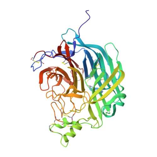
Structural and functional analyses reveal promiscuous and species specific use of ephrin receptors by Cedar virus.

Laing, E.D.Navaratnarajah, C.K.Cheliout Da Silva, S.Petzing, S.R.Xu, Y.Sterling, S.L.Marsh, G.A.Wang, L.F.Amaya, M.Nikolov, D.B.Cattaneo, R.Broder, C.C.Xu, K.

(2019) Proc Natl Acad Sci U S A 116: 20707-20715

  • DOI: https://doi.org/10.1073/pnas.1911773116
  • Primary Citation Related Structures: 
    6P72, 6P7S, 6P7Y

  • PubMed Abstract: 

    Cedar virus (CedV) is a bat-borne henipavirus related to Nipah virus (NiV) and Hendra virus (HeV), zoonotic agents of fatal human disease. CedV receptor-binding protein (G) shares only ∼30% sequence identity with those of NiV and HeV, although they can all use ephrin-B2 as an entry receptor. We demonstrate that CedV also enters cells through additional B- and A-class ephrins (ephrin-B1, ephrin-A2, and ephrin-A5) and report the crystal structure of the CedV G ectodomain alone and in complex with ephrin-B1 or ephrin-B2. The CedV G receptor-binding site is structurally distinct from other henipaviruses, underlying its capability to accommodate additional ephrin receptors. We also show that CedV can enter cells through mouse ephrin-A1 but not human ephrin-A1, which differ by 1 residue in the key contact region. This is evidence of species specific ephrin receptor usage by a henipavirus, and implicates additional ephrin receptors in potential zoonotic transmission.


  • Organizational Affiliation
    • Department of Microbiology and Immunology, Uniformed Services University of the Health Sciences, Bethesda, MD 20814.

Macromolecule Content 

  • Total Structure Weight: 140.07 kDa 
  • Atom Count: 9,852 
  • Modeled Residue Count: 1,133 
  • Deposited Residue Count: 1,148 
  • Unique protein chains: 2

Macromolecules

Find similar proteins by:|  3D Structure
Entity ID: 1
MoleculeChains  Sequence LengthOrganismDetailsImage
Attachment glycoprotein
A, C
430Cedar virusMutation(s): 0 
UniProt
Find proteins for J7H333 (Cedar virus)
Explore J7H333 
Go to UniProtKB:  J7H333
Entity Groups
Sequence Clusters30% Identity50% Identity70% Identity90% Identity95% Identity100% Identity
UniProt GroupJ7H333
Glycosylation
Glycosylation Sites: 6
Sequence Annotations
Expand
Reference Sequence
Find similar proteins by:|  3D Structure
Entity ID: 2
MoleculeChains  Sequence LengthOrganismDetailsImage
Ephrin-B2
B, D
144Homo sapiensMutation(s): 0 
Gene Names: EFNB2EPLG5HTKLLERK5
UniProt & NIH Common Fund Data Resources
Find proteins for P52799 (Homo sapiens)
Explore P52799 
Go to UniProtKB:  P52799
PHAROS:  P52799
GTEx:  ENSG00000125266 
Entity Groups
Sequence Clusters30% Identity50% Identity70% Identity90% Identity95% Identity100% Identity
UniProt GroupP52799
Sequence Annotations
Expand
Reference Sequence

Oligosaccharides

Help  
Entity ID: 3
MoleculeChains Length2D Diagram GlycosylationD Interactions
beta-D-mannopyranose-(1-4)-2-acetamido-2-deoxy-beta-D-glucopyranose-(1-4)-2-acetamido-2-deoxy-beta-D-glucopyranose
E, M
3N-Glycosylation
Glycosylation Resources
GlyTouCan: G15407YE
GlyCosmos: G15407YE
GlyGen: G15407YE
Entity ID: 4
MoleculeChains Length2D Diagram GlycosylationD Interactions
2-acetamido-2-deoxy-beta-D-glucopyranose-(1-4)-[alpha-L-fucopyranose-(1-6)]2-acetamido-2-deoxy-beta-D-glucopyranose
F, L
3N-Glycosylation
Glycosylation Resources
GlyTouCan: G21290RB
GlyCosmos: G21290RB
GlyGen: G21290RB
Entity ID: 5
MoleculeChains Length2D Diagram GlycosylationD Interactions
alpha-D-mannopyranose-(1-3)-[alpha-D-mannopyranose-(1-6)]beta-D-mannopyranose-(1-4)-2-acetamido-2-deoxy-beta-D-glucopyranose-(1-4)-[alpha-L-fucopyranose-(1-3)][alpha-L-fucopyranose-(1-6)]2-acetamido-2-deoxy-beta-D-glucopyranose
G
7N-Glycosylation
Glycosylation Resources
GlyTouCan: G11460AB
GlyCosmos: G11460AB
GlyGen: G11460AB
Entity ID: 6
MoleculeChains Length2D Diagram GlycosylationD Interactions
alpha-D-mannopyranose-(1-3)-[alpha-D-mannopyranose-(1-6)]beta-D-mannopyranose-(1-4)-2-acetamido-2-deoxy-beta-D-glucopyranose-(1-4)-2-acetamido-2-deoxy-beta-D-glucopyranose
H, I, J, K, O
5N-Glycosylation
Glycosylation Resources
GlyTouCan: G22768VO
GlyCosmos: G22768VO
GlyGen: G22768VO
Entity ID: 7
MoleculeChains Length2D Diagram GlycosylationD Interactions
alpha-D-mannopyranose-(1-6)-beta-D-mannopyranose-(1-4)-2-acetamido-2-deoxy-beta-D-glucopyranose-(1-4)-2-acetamido-2-deoxy-beta-D-glucopyranose
N
4N-Glycosylation
Glycosylation Resources
GlyTouCan: G22573RC
GlyCosmos: G22573RC
GlyGen: G22573RC

Experimental Data & Validation

Experimental Data

  • Method: X-RAY DIFFRACTION
  • Resolution: 2.84 Å
  • R-Value Free:  0.240 (Depositor), 0.242 (DCC) 
  • R-Value Work:  0.207 (Depositor), 0.208 (DCC) 
  • R-Value Observed: 0.208 (Depositor) 
Space Group: P 65
Unit Cell:
Length ( Å )Angle ( ˚ )
a = 207.021α = 90
b = 207.021β = 90
c = 120.65γ = 120
Software Package:
Software NamePurpose
HKL-2000data scaling
PHENIXrefinement
PDB_EXTRACTdata extraction
HKL-2000data reduction
PHASERphasing

Structure Validation

View Full Validation Report


Ligand Structure Quality Assessment 


Entry History 

Deposition Data

Revision History  (Full details and data files)

  • Version 1.0: 2019-09-25
    Type: Initial release
  • Version 1.1: 2019-10-09
    Changes: Data collection, Database references
  • Version 1.2: 2019-10-23
    Changes: Data collection, Database references
  • Version 2.0: 2020-07-29
    Type: Remediation
    Reason: Carbohydrate remediation
    Changes: Advisory, Atomic model, Data collection, Derived calculations, Structure summary
  • Version 2.1: 2024-11-20
    Changes: Data collection, Database references, Derived calculations, Structure summary