6P8L

Escherichia coli Bacterioferritin Substituted with Zinc Protoporphyrin IX (Zn Absorption Edge X-ray Data)


Experimental Data Snapshot

  • Method: X-RAY DIFFRACTION
  • Resolution: 2.10 Å
  • R-Value Free: 
    0.240 (Depositor), 0.240 (DCC) 
  • R-Value Work: 
    0.213 (Depositor), 0.210 (DCC) 
  • R-Value Observed: 
    0.213 (Depositor) 

Starting Model: experimental
View more details

wwPDB Validation   3D Report Full Report


Ligand Structure Quality Assessment 

Created with Raphaël 2.3.0Worse 01 BetterLigand structure goodness of fit to experimental dataBest fitted ZNHClick on this verticalbar to view details

This is version 1.1 of the entry. See complete history


Literature

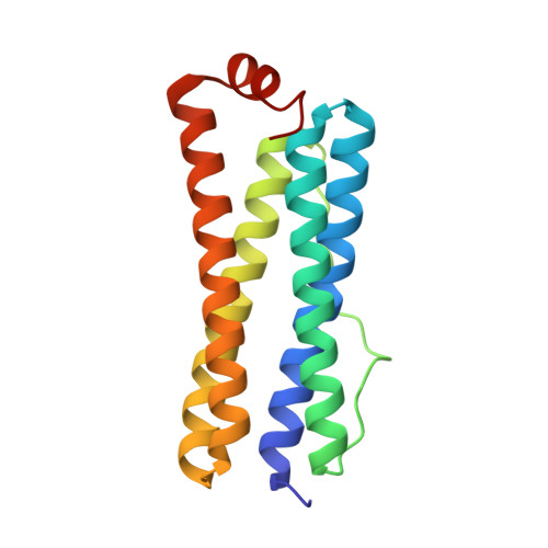
Structure of a Zinc Porphyrin-Substituted Bacterioferritin and Photophysical Properties of Iron Reduction.

Benavides, B.S.Valandro, S.Cioloboc, D.Taylor, A.B.Schanze, K.S.Kurtz Jr., D.M.

(2020) Biochemistry 59: 1618-1629

  • DOI: https://doi.org/10.1021/acs.biochem.9b01103
  • Primary Citation of Related Structures:  
    6P8K, 6P8L

  • PubMed Abstract: 

    The iron storage protein bacterioferritin (Bfr) binds up to 12 hemes b at specific sites in its protein shell. The heme b can be substituted with the photosensitizer Zn(II)-protoporphyrin IX (ZnPP), and photosensitized reductive iron release from the ferric oxyhydroxide {[FeO(OH)] n } core inside the ZnPP-Bfr protein shell was demonstrated [Cioloboc, D., et al. (2018) Biomacromolecules 19 , 178-187]. This report describes the X-ray crystal structure of ZnPP-Bfr and the effects of loaded iron on the photophysical properties of the ZnPP. The crystal structure of ZnPP-Bfr shows a unique six-coordinate zinc in the ZnPP with two axial methionine sulfur ligands. Steady state and transient ultraviolet-visible absorption and luminescence spectroscopies show that irradiation with light overlapping the Soret absorption causes oxidation of ZnPP to the cation radical ZnPP •+ only when the ZnPP-Bfr is loaded with [FeO(OH)] n . Femtosecond transient absorption spectroscopy shows that this photooxidation occurs from the singlet excited state ( 1 ZnPP*) on the picosecond time scale and is consistent with two oxidizing populations of Fe 3+ , which do not appear to involve the ferroxidase center iron. We propose that [FeO(OH)] n clusters at or near the inner surface of the protein shell are responsible for ZnPP photooxidation. Hopping of the photoinjected electrons through the [FeO(OH)] n would effectively cause migration of Fe 2+ through the inner cavity to pores where it exits the protein. Reductive iron mobilization is presumed to be a physiological function of Bfrs. The phototriggered Fe 3+ reduction could be used to identify the sites of iron mobilization within the Bfr protein shell.


  • Organizational Affiliation

    Department of Chemistry, University of Texas at San Antonio, San Antonio, Texas 78249, United States.


Macromolecules
Find similar proteins by:  (by identity cutoff)  |  3D Structure
Entity ID: 1
MoleculeChains Sequence LengthOrganismDetailsImage
Bacterioferritin
A, B, C, D, E
158Escherichia coli K-12Mutation(s): 0 
Gene Names: bfrb3336JW3298
EC: 1.16.3.1
UniProt
Find proteins for P0ABD3 (Escherichia coli (strain K12))
Explore P0ABD3 
Go to UniProtKB:  P0ABD3
Entity Groups  
Sequence Clusters30% Identity50% Identity70% Identity90% Identity95% Identity100% Identity
UniProt GroupP0ABD3
Sequence Annotations
Expand
  • Reference Sequence
Small Molecules
Ligands 4 Unique
IDChains Name / Formula / InChI Key2D Diagram3D Interactions
ZNH (Subject of Investigation/LOI)
Query on ZNH

Download Ideal Coordinates CCD File 
AA [auth D]
GB [auth L]
IA [auth F]
R [auth B]
RA [auth H]
PROTOPORPHYRIN IX CONTAINING ZN
C34 H32 N4 O4 Zn
FUTVBRXUIKZACV-RGGAHWMASA-L
MLI
Query on MLI

Download Ideal Coordinates CCD File 
AB [auth J]
BB [auth J]
CB [auth K]
DA [auth D]
EA [auth D]
MALONATE ION
C3 H2 O4
OFOBLEOULBTSOW-UHFFFAOYSA-L
ZN
Query on ZN

Download Ideal Coordinates CCD File 
BA [auth D]
DB [auth K]
FA [auth E]
HB [auth L]
JA [auth F]
ZINC ION
Zn
PTFCDOFLOPIGGS-UHFFFAOYSA-N
NA
Query on NA

Download Ideal Coordinates CCD File 
CA [auth D],
KA [auth F],
N [auth A],
T [auth B]
SODIUM ION
Na
FKNQFGJONOIPTF-UHFFFAOYSA-N
Experimental Data & Validation

Experimental Data

  • Method: X-RAY DIFFRACTION
  • Resolution: 2.10 Å
  • R-Value Free:  0.240 (Depositor), 0.240 (DCC) 
  • R-Value Work:  0.213 (Depositor), 0.210 (DCC) 
  • R-Value Observed: 0.213 (Depositor) 
Space Group: P 42 21 2
Unit Cell:
Length ( Å )Angle ( ˚ )
a = 207.918α = 90
b = 207.918β = 90
c = 143.046γ = 90
Software Package:
Software NamePurpose
PHENIXrefinement
XDSdata reduction
XDSdata scaling
PHENIXphasing

Structure Validation

View Full Validation Report


Ligand Structure Quality Assessment 

Created with Raphaël 2.3.0Worse 01 BetterLigand structure goodness of fit to experimental dataBest fitted ZNHClick on this verticalbar to view details

Entry History & Funding Information

Deposition Data


Funding OrganizationLocationGrant Number
National Institutes of Health/National Center for Advancing Translational Sciences (NIH/NCATS)United States8UL1TR001120

Revision History  (Full details and data files)

  • Version 1.0: 2020-05-13
    Type: Initial release
  • Version 1.1: 2023-10-11
    Changes: Data collection, Database references, Derived calculations, Refinement description