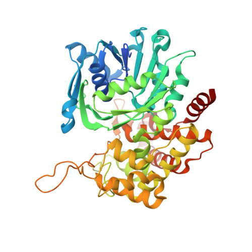
6PBP

Pseudopaline Dehydrogenase with (S)-Pseudopaline Soaked 1 hour


Experimental Data Snapshot

  • Method: X-RAY DIFFRACTION
  • Resolution: 1.64 Å
  • R-Value Free: 0.201 
  • R-Value Work: 0.169 
  • R-Value Observed: 0.170 

Starting Model: experimental
View more details

wwPDB Validation   3D Report Full Report


Ligand Structure Quality Assessment 


This is version 1.3 of the entry. See complete history


Literature

Staphylopine and pseudopaline dehydrogenase from bacterial pathogens catalyze reversible reactions and produce stereospecific metallophores.

McFarlane, J.S.Zhang, J.Wang, S.Lei, X.Moran, G.R.Lamb, A.L.

(2019) J Biol Chem 294: 17988-18001

  • DOI: https://doi.org/10.1074/jbc.RA119.011059
  • Primary Citation of Related Structures:  
    6PBM, 6PBN, 6PBP, 6PBT

  • PubMed Abstract: 

    Pseudopaline and staphylopine are opine metallophores biosynthesized by Pseudomonas aeruginosa and Staphylococcus aureus , respectively. The final step in opine metallophore biosynthesis is the condensation of the product of a nicotianamine (NA) synthase reaction ( i.e. l-HisNA for pseudopaline and d-HisNA for staphylopine) with an α-keto acid (α-ketoglutarate for pseudopaline and pyruvate for staphylopine), which is performed by an opine dehydrogenase. We hypothesized that the opine dehydrogenase reaction would be reversible only for the opine metallophore product with ( R )-stereochemistry at carbon C2 of the α-keto acid (prochiral prior to catalysis). A kinetic analysis using stopped-flow spectrometry with ( R )- or ( S )-staphylopine and kinetic and structural analysis with ( R )- and ( S )-pseudopaline confirmed catalysis in the reverse direction for only ( R )-staphylopine and ( R )-pseudopaline, verifying the stereochemistry of these two opine metallophores. Structural analysis at 1.57-1.85 Å resolution captured the hydrolysis of ( R )-pseudopaline and allowed identification of a binding pocket for the l-histidine moiety of pseudopaline formed through a repositioning of Phe-340 and Tyr-289 during the catalytic cycle. Transient-state kinetic analysis revealed an ordered release of NADP + followed by staphylopine, with staphylopine release being the rate-limiting step in catalysis. Knowledge of the stereochemistry for opine metallophores has implications for future studies involving kinetic analysis, as well as opine metallophore transport, metal coordination, and the generation of chiral amines for pharmaceutical development.


  • Organizational Affiliation

    Department of Molecular Biosciences, University of Kansas, Lawrence, Kansas 66045.


Macromolecules
Find similar proteins by:  (by identity cutoff)  |  3D Structure
Entity ID: 1
MoleculeChains Sequence LengthOrganismDetailsImage
Pseudopaline Dehydrogenase
A, B
449Pseudomonas aeruginosa PAO1Mutation(s): 0 
Gene Names: PA4835
EC: 1.5.1
UniProt
Find proteins for Q9HUX5 (Pseudomonas aeruginosa (strain ATCC 15692 / DSM 22644 / CIP 104116 / JCM 14847 / LMG 12228 / 1C / PRS 101 / PAO1))
Explore Q9HUX5 
Go to UniProtKB:  Q9HUX5
Entity Groups  
Sequence Clusters30% Identity50% Identity70% Identity90% Identity95% Identity100% Identity
UniProt GroupQ9HUX5
Sequence Annotations
Expand
  • Reference Sequence
Small Molecules
Ligands 3 Unique
IDChains Name / Formula / InChI Key2D Diagram3D Interactions
NAP (Subject of Investigation/LOI)
Query on NAP

Download Ideal Coordinates CCD File 
C [auth A],
K [auth B]
NADP NICOTINAMIDE-ADENINE-DINUCLEOTIDE PHOSPHATE
C21 H28 N7 O17 P3
XJLXINKUBYWONI-NNYOXOHSSA-N
O7J (Subject of Investigation/LOI)
Query on O7J

Download Ideal Coordinates CCD File 
D [auth A],
L [auth B]
N-[(1S)-1-carboxy-3-{[(1S)-1-carboxy-2-(1H-imidazol-5-yl)ethyl]amino}propyl]-L-glutamic acid
C15 H22 N4 O8
RLSXUJSGKUUKFH-DCAQKATOSA-N
EDO
Query on EDO

Download Ideal Coordinates CCD File 
E [auth A]
F [auth A]
G [auth A]
H [auth A]
I [auth A]
E [auth A],
F [auth A],
G [auth A],
H [auth A],
I [auth A],
J [auth A],
M [auth B],
N [auth B]
1,2-ETHANEDIOL
C2 H6 O2
LYCAIKOWRPUZTN-UHFFFAOYSA-N
Experimental Data & Validation

Experimental Data

  • Method: X-RAY DIFFRACTION
  • Resolution: 1.64 Å
  • R-Value Free: 0.201 
  • R-Value Work: 0.169 
  • R-Value Observed: 0.170 
  • Space Group: C 1 2 1
Unit Cell:
Length ( Å )Angle ( ˚ )
a = 180.633α = 90
b = 53.758β = 98.69
c = 96.802γ = 90
Software Package:
Software NamePurpose
PHENIXrefinement
XDSdata reduction
XDSdata scaling
PHASERphasing

Structure Validation

View Full Validation Report


Ligand Structure Quality Assessment 


Entry History & Funding Information

Deposition Data


Funding OrganizationLocationGrant Number
National Institutes of Health/National Institute of General Medical Sciences (NIH/NIGMS)United StatesR01-GM127655-01
National Institutes of Health/National Institute of General Medical Sciences (NIH/NIGMS)United StatesP20 GM103418
American Heart AssociationUnited StatesPRE33960374
National Institutes of Health/National Institute of General Medical Sciences (NIH/NIGMS)United StatesT32-GM08545

Revision History  (Full details and data files)

  • Version 1.0: 2019-10-30
    Type: Initial release
  • Version 1.1: 2019-12-04
    Changes: Database references
  • Version 1.2: 2020-01-01
    Changes: Author supporting evidence
  • Version 1.3: 2023-10-11
    Changes: Data collection, Database references, Refinement description