6PVI

Crystal structure of PhqK in complex with paraherquamide L


Experimental Data Snapshot

  • Method: X-RAY DIFFRACTION
  • Resolution: 2.09 Å
  • R-Value Free: 0.256 
  • R-Value Work: 0.208 
  • R-Value Observed: 0.210 

wwPDB Validation   3D Report Full Report


Ligand Structure Quality Assessment 


This is version 1.2 of the entry. See complete history


Literature

Molecular Basis for Spirocycle Formation in the Paraherquamide Biosynthetic Pathway.

Fraley, A.E.Caddell Haatveit, K.Ye, Y.Kelly, S.P.Newmister, S.A.Yu, F.Williams, R.M.Smith, J.L.Houk, K.N.Sherman, D.H.

(2020) J Am Chem Soc 142: 2244-2252

  • DOI: https://doi.org/10.1021/jacs.9b09070
  • Primary Citation of Related Structures:  
    6PVF, 6PVG, 6PVH, 6PVI, 6PVJ

  • PubMed Abstract: 

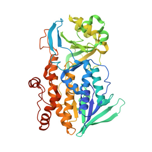
    The paraherquamides are potent anthelmintic natural products with complex heptacyclic scaffolds. One key feature of these molecules is the spiro-oxindole moiety that lends a strained three-dimensional architecture to these structures. The flavin monooxygenase PhqK was found to catalyze spirocycle formation through two parallel pathways in the biosynthesis of paraherquamides A and G. Two new paraherquamides (K and L) were isolated from a Δ phqK strain of Penicillium simplicissimum , and subsequent enzymatic reactions with these compounds generated two additional metabolites, paraherquamides M and N. Crystal structures of PhqK in complex with various substrates provided a foundation for mechanistic analyses and computational studies. While it is evident that PhqK can react with various substrates, reaction kinetics and molecular dynamics simulations indicated that the dioxepin-containing paraherquamide L is the favored substrate. Through this effort, we have elucidated a key step in the biosynthesis of the paraherquamides and provided a rationale for the selective spirocyclization of these powerful anthelmintic agents.


  • Organizational Affiliation

    Department of Chemistry and Biochemistry , University of California , Los Angeles , California 90095 , United States.


Macromolecules
Find similar proteins by:  (by identity cutoff)  |  3D Structure
Entity ID: 1
MoleculeChains Sequence LengthOrganismDetailsImage
FAD monooxygenase459Penicillium fellutanumMutation(s): 0 
Gene Names: phqK
UniProt
Find proteins for L0E4H0 (Penicillium fellutanum)
Explore L0E4H0 
Go to UniProtKB:  L0E4H0
Entity Groups  
Sequence Clusters30% Identity50% Identity70% Identity90% Identity95% Identity100% Identity
UniProt GroupL0E4H0
Sequence Annotations
Expand
  • Reference Sequence
Small Molecules
Ligands 2 Unique
IDChains Name / Formula / InChI Key2D Diagram3D Interactions
FAD (Subject of Investigation/LOI)
Query on FAD

Download Ideal Coordinates CCD File 
C [auth A]FLAVIN-ADENINE DINUCLEOTIDE
C27 H33 N9 O15 P2
VWWQXMAJTJZDQX-UYBVJOGSSA-N
OZ7 (Subject of Investigation/LOI)
Query on OZ7

Download Ideal Coordinates CCD File 
B [auth A](8aS,13S,13aR,14aS)-4,4,13,15,15-pentamethyl-12,13,14,14a,15,16-hexahydro-4H,8H,9H,11H-8a,13a-(epiminomethano)[1,4]dioxepino[2,3-a]indolizino[6,7-h]carbazol-17-one
C27 H33 N3 O3
CHODRHYJOPXZLV-ZYHIPPEZSA-N
Experimental Data & Validation

Experimental Data

  • Method: X-RAY DIFFRACTION
  • Resolution: 2.09 Å
  • R-Value Free: 0.256 
  • R-Value Work: 0.208 
  • R-Value Observed: 0.210 
  • Space Group: P 21 21 21
Unit Cell:
Length ( Å )Angle ( ˚ )
a = 64.903α = 90
b = 79.91β = 90
c = 87.687γ = 90
Software Package:
Software NamePurpose
PHENIXrefinement

Structure Validation

View Full Validation Report


Ligand Structure Quality Assessment 


Entry History & Funding Information

Deposition Data


Funding OrganizationLocationGrant Number
National Institutes of Health/National Human Genome Research Institute (NIH/NHGRI)United StatesR01 CA70375

Revision History  (Full details and data files)

  • Version 1.0: 2020-01-22
    Type: Initial release
  • Version 1.1: 2020-02-19
    Changes: Database references
  • Version 1.2: 2024-03-13
    Changes: Data collection, Database references