6PVL | pdb_00006pvl

Cryo-EM structure of mouse TRPV3 in closed state at 42 degrees Celsius


Experimental Data Snapshot

  • Method: ELECTRON MICROSCOPY
  • Resolution: 4.40 Å
  • Aggregation State: PARTICLE 
  • Reconstruction Method: SINGLE PARTICLE 

wwPDB Validation 3D Report Full Report

Validation slider image for 6PVL

This is version 1.4 of the entry. See complete history

Literature

Structural basis of temperature sensation by the TRP channel TRPV3.

Singh, A.K.McGoldrick, L.L.Demirkhanyan, L.Leslie, M.Zakharian, E.Sobolevsky, A.I.

(2019) Nat Struct Mol Biol 26: 994-998

  • DOI: https://doi.org/10.1038/s41594-019-0318-7
  • Primary Citation Related Structures: 
    6PVL, 6PVM, 6PVN, 6PVO, 6PVP, 6PVQ

  • PubMed Abstract: 

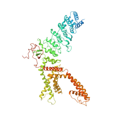
    We present structures of mouse TRPV3 in temperature-dependent open, closed and intermediate states that suggest two-step activation of TRPV3 by heat. During the strongly temperature-dependent first step, sensitization, the channel pore remains closed while S6 helices undergo α-to-π transitions. During the weakly temperature-dependent second step, channel opening, tight association of the S1-S4 and pore domains is stabilized by changes in the carboxy-terminal and linker domains.


  • Organizational Affiliation
    • Department of Biochemistry and Molecular Biophysics, Columbia University, New York, NY, USA.

Macromolecule Content 

  • Total Structure Weight: 370.57 kDa 
  • Atom Count: 21,006 
  • Modeled Residue Count: 2,576 
  • Deposited Residue Count: 3,232 
  • Unique protein chains: 1

Macromolecules

Find similar proteins by:|  3D Structure
Entity ID: 1
MoleculeChains  Sequence LengthOrganismDetailsImage
Transient receptor potential cation channel subfamily V member 3
A, B, C, D
808Mus musculusMutation(s): 0 
Gene Names: Trpv3
Membrane Entity: Yes 
UniProt
Find proteins for Q8K424 (Mus musculus)
Explore Q8K424 
Go to UniProtKB:  Q8K424
Entity Groups
Sequence Clusters30% Identity50% Identity70% Identity90% Identity95% Identity100% Identity
UniProt GroupQ8K424
Sequence Annotations
Expand
Reference Sequence

Experimental Data & Validation

Experimental Data

  • Method: ELECTRON MICROSCOPY
  • Resolution: 4.40 Å
  • Aggregation State: PARTICLE 
  • Reconstruction Method: SINGLE PARTICLE 
EM Software:
TaskSoftware PackageVersion
RECONSTRUCTIONRELION

Structure Validation

View Full Validation Report


Entry History 

& Funding Information

Deposition Data


Funding OrganizationLocationGrant Number
National Institutes of Health/National Cancer Institute (NIH/NCI)United StatesR01 CA206573

Revision History  (Full details and data files)

  • Version 1.0: 2019-10-23
    Type: Initial release
  • Version 1.1: 2019-11-06
    Changes: Data collection, Database references
  • Version 1.2: 2019-11-27
    Changes: Database references
  • Version 1.3: 2019-12-04
    Changes: Author supporting evidence
  • Version 1.4: 2024-03-20
    Changes: Data collection, Database references