6Q1U

Structure of plasmin and peptide complex


Experimental Data Snapshot

  • Method: X-RAY DIFFRACTION
  • Resolution: 2.35 Å
  • R-Value Free: 0.287 
  • R-Value Work: 0.233 
  • R-Value Observed: 0.235 

wwPDB Validation   3D Report Full Report


Ligand Structure Quality Assessment 


This is version 1.3 of the entry. See complete history


Literature

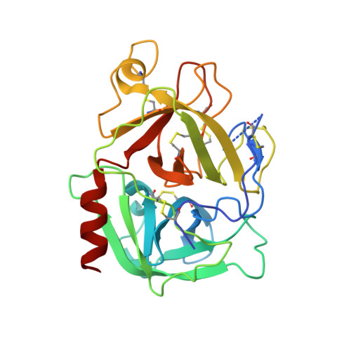
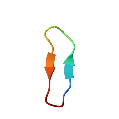
Application and Structural Analysis of Triazole-Bridged Disulfide Mimetics in Cyclic Peptides.

White, A.M.de Veer, S.J.Wu, G.Harvey, P.J.Yap, K.King, G.J.Swedberg, J.E.Wang, C.K.Law, R.H.P.Durek, T.Craik, D.J.

(2020) Angew Chem Int Ed Engl 59: 11273-11277

  • DOI: https://doi.org/10.1002/anie.202003435
  • Primary Citation of Related Structures:  
    6Q1U, 6U22, 6U24, 6U7Q, 6U7R, 6U7S, 6U7U, 6U7W, 6U7X, 6VXY, 6VY8

  • PubMed Abstract: 

    Ruthenium-catalysed azide-alkyne cycloaddition (RuAAC) provides access to 1,5-disubstituted 1,2,3-triazole motifs in peptide engineering applications. However, investigation of this motif as a disulfide mimetic in cyclic peptides has been limited, and the structural consequences remain to be studied. We report synthetic strategies to install various triazole linkages into cyclic peptides through backbone cyclisation and RuAAC cross-linking reactions. These linkages were evaluated in four serine protease inhibitors based on sunflower trypsin inhibitor-1. NMR and X-ray crystallography revealed exceptional consensus of bridging distance and backbone conformations (RMSD<0.5 Å) of the triazole linkages compared to the parent disulfide molecules. The triazole-bridged peptides also displayed superior half-lives in liver S9 stability assays compared to disulfide-bridged peptides. This work establishes a foundation for the application of 1,5-disubstituted 1,2,3-triazoles as disulfide mimetics.


  • Organizational Affiliation

    ARC Centre of Excellence for Innovations in Peptide and Protein Science, Institute for Molecular Bioscience, The University of Queensland, Brisbane, QLD, 4072, Australia.


Macromolecules
Find similar proteins by:  (by identity cutoff)  |  3D Structure
Entity ID: 1
MoleculeChains Sequence LengthOrganismDetailsImage
Plasminogen
A, B
249Homo sapiensMutation(s): 0 
Gene Names: PLG
EC: 3.4.21.7
UniProt & NIH Common Fund Data Resources
Find proteins for P00747 (Homo sapiens)
Explore P00747 
Go to UniProtKB:  P00747
PHAROS:  P00747
GTEx:  ENSG00000122194 
Entity Groups  
Sequence Clusters30% Identity50% Identity70% Identity90% Identity95% Identity100% Identity
UniProt GroupP00747
Sequence Annotations
Expand
  • Reference Sequence

Find similar proteins by:  Sequence   |   3D Structure  

Entity ID: 2
MoleculeChains Sequence LengthOrganismDetailsImage
GLY-ARG-ALA-TYR-LYS-SER-LYS-PRO-PRO-ILE-ALA-PHE-PRO-ASP
C, D
14Helianthus annuusMutation(s): 0 
UniProt
Find proteins for Q4GWU5 (Helianthus annuus)
Explore Q4GWU5 
Go to UniProtKB:  Q4GWU5
Entity Groups  
Sequence Clusters30% Identity50% Identity70% Identity90% Identity95% Identity100% Identity
UniProt GroupQ4GWU5
Sequence Annotations
Expand
  • Reference Sequence
Experimental Data & Validation

Experimental Data

  • Method: X-RAY DIFFRACTION
  • Resolution: 2.35 Å
  • R-Value Free: 0.287 
  • R-Value Work: 0.233 
  • R-Value Observed: 0.235 
  • Space Group: P 21 21 21
Unit Cell:
Length ( Å )Angle ( ˚ )
a = 79.228α = 90
b = 80.293β = 90
c = 83.198γ = 90
Software Package:
Software NamePurpose
PHENIXrefinement
XDSdata reduction
SCALAdata scaling
PHASERphasing

Structure Validation

View Full Validation Report


Ligand Structure Quality Assessment 


Entry History 

Deposition Data

Revision History  (Full details and data files)

  • Version 1.0: 2020-04-15
    Type: Initial release
  • Version 1.1: 2020-04-22
    Changes: Database references
  • Version 1.2: 2020-07-15
    Changes: Database references
  • Version 1.3: 2023-10-11
    Changes: Data collection, Database references, Derived calculations, Refinement description