


Crystal structure of the O-GlcNAc transferase Asn648Tyr mutation
Gundogdu, M., van Aalten, D.M.F.To be published.
Experimental Data Snapshot
Entity ID: 1 | |||||
|---|---|---|---|---|---|
| Molecule | Chains | Sequence Length | Organism | Details | Image |
| UDP-N-acetylglucosamine--peptide N-acetylglucosaminyltransferase 110 kDa subunit | 723 | Homo sapiens | Mutation(s): 1 Gene Names: OGT EC: 2.4.1.255 | 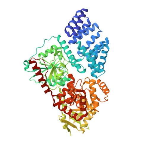 | |
UniProt & NIH Common Fund Data Resources | |||||
PHAROS: O15294 GTEx: ENSG00000147162 | |||||
Entity Groups | |||||
| Sequence Clusters | 30% Identity50% Identity70% Identity90% Identity95% Identity100% Identity | ||||
| UniProt Group | O15294 | ||||
Sequence AnnotationsExpand | |||||
Reference Sequence | |||||
| Ligands 2 Unique | |||||
|---|---|---|---|---|---|
| ID | Chains | Name / Formula / InChI Key | 2D Diagram | 3D Interactions | |
| 12V Download:Ideal Coordinates CCD File | B [auth A] | (2S,3R,4R,5S,6R)-3-(acetylamino)-4,5-dihydroxy-6-(hydroxymethyl)tetrahydro-2H-thiopyran-2-yl [(2R,3S,4R,5R)-5-(2,4-dioxo-3,4-dihydropyrimidin-1(2H)-yl)-3,4-dihydroxytetrahydrofuran-2-yl]methyl dihydrogen diphosphate C17 H27 N3 O16 P2 S JPRVHSQHWXZSNC-UBDZBXRQSA-N |  | ||
| PO4 (Subject of Investigation/LOI) Download:Ideal Coordinates CCD File | C [auth A], D [auth A] | PHOSPHATE ION O4 P NBIIXXVUZAFLBC-UHFFFAOYSA-K |  | ||
| Length ( Å ) | Angle ( ˚ ) |
|---|---|
| a = 137.316 | α = 90 |
| b = 150.736 | β = 90 |
| c = 199.492 | γ = 90 |
| Software Name | Purpose |
|---|---|
| REFMAC | refinement |
| MOSFLM | data reduction |
| SCALA | data scaling |
| MOLREP | phasing |
| Funding Organization | Location | Grant Number |
|---|---|---|
| Wellcome Trust | United Kingdom | 110061 |


