6Q9O

HDM2 (17-111, WILDTYPE) COMPLEXED WITH COMPOUND 10 AT 1.21A; Structural states of Hdm2 and HdmX: X-ray elucidation of adaptations and binding interactions for different chemical compound classes


Experimental Data Snapshot

  • Method: X-RAY DIFFRACTION
  • Resolution: 1.21 Å
  • R-Value Free: 0.156 
  • R-Value Work: 0.145 
  • R-Value Observed: 0.145 

wwPDB Validation   3D Report Full Report


Ligand Structure Quality Assessment 


This is version 1.2 of the entry. See complete history


Literature

Structural States of Hdm2 and HdmX: X-ray Elucidation of Adaptations and Binding Interactions for Different Chemical Compound Classes.

Kallen, J.Izaac, A.Chau, S.Wirth, E.Schoepfer, J.Mah, R.Schlapbach, A.Stutz, S.Vaupel, A.Guagnano, V.Masuya, K.Stachyra, T.M.Salem, B.Chene, P.Gessier, F.Holzer, P.Furet, P.

(2019) ChemMedChem 14: 1305-1314

  • DOI: https://doi.org/10.1002/cmdc.201900201
  • Primary Citation of Related Structures:  
    6Q96, 6Q9H, 6Q9L, 6Q9O, 6Q9Q, 6Q9S, 6Q9U, 6Q9W, 6Q9Y

  • PubMed Abstract: 

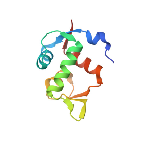
    Hdm2 (human MDM2, human double minute 2 homologue) counteracts p53 function by direct binding to p53 and by ubiquitin-dependent p53 protein degradation. Activation of p53 by inhibitors of the p53-Hdm2 interaction is being pursued as a therapeutic strategy in p53 wild-type cancers. In addition, HdmX (human MDMX, human MDM4) was also identified as an important therapeutic target to efficiently reactivate p53, and it is likely that dual inhibition of Hdm2 and HdmX is beneficial. Herein we report four new X-ray structures for Hdm2 and five new X-ray structures for HdmX complexes, involving different classes of synthetic compounds (including the worldwide highest resolutions for Hdm2 and HdmX, at 1.13 and 1.20 Å, respectively). We also reveal the key additive 18-crown-ether, which we discovered to enable HdmX crystallization and show its stabilization of various Lys residues. In addition, we report the previously unpublished details of X-ray structure determinations for eight further Hdm2 complexes, including the clinical trial compounds NVP-CGM097 and NVP-HDM201. An analysis of all compound binding modes reveals new and deepened insight into the possible adaptations and structural states of Hdm2 (e.g., flip of F55, flip of Y67, reorientation of H96) and HdmX (e.g., flip of H55, dimer induction), enabling key binding interactions for different compound classes. To facilitate comparisons, we used the same numbering for Hdm2 (as in Q00987) and HdmX (as in O15151, but minus 1). Taken together, these structural insights should prove useful for the design and optimization of further selective and/or dual Hdm2/HdmX inhibitors.


  • Organizational Affiliation

    Chemical Biology & Therapeutics, Novartis Institutes for BioMedical Research, Novartis Campus, 4002, Basel, Switzerland.


Macromolecules
Find similar proteins by:  (by identity cutoff)  |  3D Structure
Entity ID: 1
MoleculeChains Sequence LengthOrganismDetailsImage
E3 ubiquitin-protein ligase Mdm2
A, B
96Homo sapiensMutation(s): 0 
Gene Names: MDM2
EC: 2.3.2.27
UniProt & NIH Common Fund Data Resources
Find proteins for Q00987 (Homo sapiens)
Explore Q00987 
Go to UniProtKB:  Q00987
PHAROS:  Q00987
GTEx:  ENSG00000135679 
Entity Groups  
Sequence Clusters30% Identity50% Identity70% Identity90% Identity95% Identity100% Identity
UniProt GroupQ00987
Sequence Annotations
Expand
  • Reference Sequence
Small Molecules
Ligands 1 Unique
IDChains Name / Formula / InChI Key2D Diagram3D Interactions
HU8 (Subject of Investigation/LOI)
Query on HU8

Download Ideal Coordinates CCD File 
C [auth A],
D [auth B]
~{N}-~{tert}-butyl-2-[4-chloranyl-2-[5-(3-chloranyl-4-fluoranyl-phenyl)-2-cyclohexyl-4-(1~{H}-1,2,3,4-tetrazol-5-yl)imidazol-1-yl]phenyl]ethanamide
C28 H30 Cl2 F N7 O
SFBXQXHCYMXGGU-UHFFFAOYSA-N
Binding Affinity Annotations 
IDSourceBinding Affinity
HU8 Binding MOAD:  6Q9O IC50: 3.4 (nM) from 1 assay(s)
Experimental Data & Validation

Experimental Data

  • Method: X-RAY DIFFRACTION
  • Resolution: 1.21 Å
  • R-Value Free: 0.156 
  • R-Value Work: 0.145 
  • R-Value Observed: 0.145 
  • Space Group: P 1 21 1
Unit Cell:
Length ( Å )Angle ( ˚ )
a = 37.929α = 90
b = 58.976β = 106.82
c = 39.88γ = 90
Software Package:
Software NamePurpose
DENZOdata reduction
SCALEPACKdata scaling
PHASERphasing
REFMACrefinement
PDB_EXTRACTdata extraction

Structure Validation

View Full Validation Report


Ligand Structure Quality Assessment 


Entry History 

Deposition Data

  • Released Date: 2019-05-15 
  • Deposition Author(s): Kallen, J.

Revision History  (Full details and data files)

  • Version 1.0: 2019-05-15
    Type: Initial release
  • Version 1.1: 2019-07-24
    Changes: Data collection, Database references
  • Version 1.2: 2024-01-24
    Changes: Data collection, Database references, Refinement description