6QGT

The carbon monoxide inhibition of F420-reducing [NiFe] hydrogenase complex from Methanosarcina barkeri


Experimental Data Snapshot

  • Method: X-RAY DIFFRACTION
  • Resolution: 1.99 Å
  • R-Value Free: 
    0.209 (Depositor), 0.210 (DCC) 
  • R-Value Work: 
    0.169 (Depositor), 0.170 (DCC) 
  • R-Value Observed: 
    0.170 (Depositor) 

Starting Model: experimental
View more details

wwPDB Validation   3D Report Full Report


Ligand Structure Quality Assessment 

Created with Raphaël 2.3.0Worse 01 BetterLigand structure goodness of fit to experimental dataBest fitted FADClick on this verticalbar to view details

This is version 1.3 of the entry. See complete history


Literature

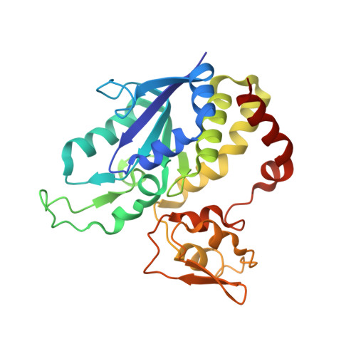
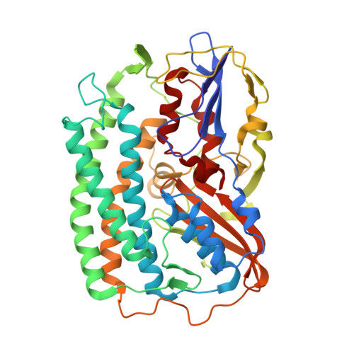
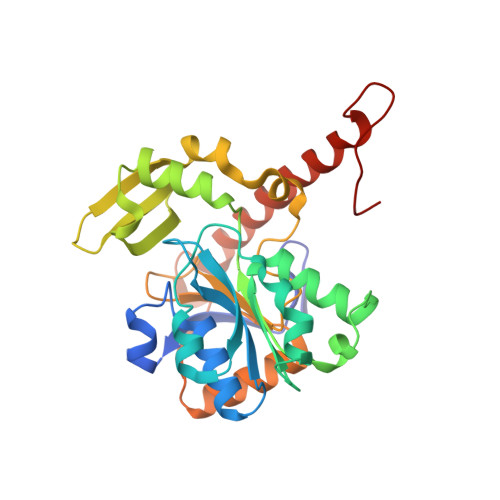
X-ray Crystallography and Vibrational Spectroscopy Reveal the Key Determinants of Biocatalytic Dihydrogen Cycling by [NiFe] Hydrogenases.

Ilina, Y.Lorent, C.Katz, S.Jeoung, J.H.Shima, S.Horch, M.Zebger, I.Dobbek, H.

(2019) Angew Chem Int Ed Engl 58: 18710-18714

  • DOI: https://doi.org/10.1002/anie.201908258
  • Primary Citation of Related Structures:  
    6QGR, 6QGT, 6QII

  • PubMed Abstract: 

    [NiFe] hydrogenases are complex model enzymes for the reversible cleavage of dihydrogen (H 2 ). However, structural determinants of efficient H 2 binding to their [NiFe] active site are not properly understood. Here, we present crystallographic and vibrational-spectroscopic insights into the unexplored structure of the H 2 -binding [NiFe] intermediate. Using an F 420 -reducing [NiFe]-hydrogenase from Methanosarcina barkeri as a model enzyme, we show that the protein backbone provides a strained chelating scaffold that tunes the [NiFe] active site for efficient H 2 binding and conversion. The protein matrix also directs H 2 diffusion to the [NiFe] site via two gas channels and allows the distribution of electrons between functional protomers through a subunit-bridging FeS cluster. Our findings emphasize the relevance of an atypical Ni coordination, thereby providing a blueprint for the design of bio-inspired H 2 -conversion catalysts.


  • Organizational Affiliation

    Institut für Biologie, Strukturbiologie/Biochemie, Humboldt-Universität zu Berlin, Philippstraße 13, 10115, Berlin, Germany.


Macromolecules
Find similar proteins by:  (by identity cutoff)  |  3D Structure
Entity ID: 1
MoleculeChains Sequence LengthOrganismDetailsImage
Coenzyme F420 hydrogenase subunit gammaA [auth G]253Methanosarcina barkeri MSMutation(s): 0 
EC: 1.12.98.1
UniProt
Find proteins for A0A0E3LP72 (Methanosarcina barkeri MS)
Explore A0A0E3LP72 
Go to UniProtKB:  A0A0E3LP72
Entity Groups  
Sequence Clusters30% Identity50% Identity70% Identity90% Identity95% Identity100% Identity
UniProt GroupA0A0E3LP72
Sequence Annotations
Expand
  • Reference Sequence
Find similar proteins by:  (by identity cutoff)  |  3D Structure
Entity ID: 2
MoleculeChains Sequence LengthOrganismDetailsImage
F420-reducing hydrogenase, subunit alphaB [auth A]438Methanosarcina barkeri MSMutation(s): 0 
EC: 1.12.98.1
UniProt
Find proteins for A0A0E3QYL7 (Methanosarcina barkeri MS)
Explore A0A0E3QYL7 
Go to UniProtKB:  A0A0E3QYL7
Entity Groups  
Sequence Clusters30% Identity50% Identity70% Identity90% Identity95% Identity100% Identity
UniProt GroupA0A0E3QYL7
Sequence Annotations
Expand
  • Reference Sequence
Find similar proteins by:  (by identity cutoff)  |  3D Structure
Entity ID: 3
MoleculeChains Sequence LengthOrganismDetailsImage
Coenzyme F420 hydrogenase subunit betaC [auth B]291Methanosarcina barkeri MSMutation(s): 0 
EC: 1.12.98.1
UniProt
Find proteins for A0A0E3QWH3 (Methanosarcina barkeri MS)
Explore A0A0E3QWH3 
Go to UniProtKB:  A0A0E3QWH3
Entity Groups  
Sequence Clusters30% Identity50% Identity70% Identity90% Identity95% Identity100% Identity
UniProt GroupA0A0E3QWH3
Sequence Annotations
Expand
  • Reference Sequence
Small Molecules
Ligands 9 Unique
IDChains Name / Formula / InChI Key2D Diagram3D Interactions
FAD
Query on FAD

Download Ideal Coordinates CCD File 
Q [auth B]FLAVIN-ADENINE DINUCLEOTIDE
C27 H33 N9 O15 P2
VWWQXMAJTJZDQX-UYBVJOGSSA-N
SF4
Query on SF4

Download Ideal Coordinates CCD File 
D [auth G],
E [auth G],
F [auth G],
R [auth B]
IRON/SULFUR CLUSTER
Fe4 S4
LJBDFODJNLIPKO-UHFFFAOYSA-N
J52
Query on J52

Download Ideal Coordinates CCD File 
K [auth A]dicyano-(oxidaniumylidynemethylnickelio)-(oxidanylidenemethylidene)iron
C4 Fe N2 Ni O2
MJFCHCXJXOCHFY-UHFFFAOYSA-N
FES
Query on FES

Download Ideal Coordinates CCD File 
G
FE2/S2 (INORGANIC) CLUSTER
Fe2 S2
NIXDOXVAJZFRNF-UHFFFAOYSA-N
144
Query on 144

Download Ideal Coordinates CCD File 
H [auth G],
N [auth A]
TRIS-HYDROXYMETHYL-METHYL-AMMONIUM
C4 H12 N O3
DRDCQJADRSJFFD-UHFFFAOYSA-N
MPD
Query on MPD

Download Ideal Coordinates CCD File 
O [auth A](4S)-2-METHYL-2,4-PENTANEDIOL
C6 H14 O2
SVTBMSDMJJWYQN-YFKPBYRVSA-N
BU3
Query on BU3

Download Ideal Coordinates CCD File 
I [auth G],
J [auth G],
P [auth B]
(R,R)-2,3-BUTANEDIOL
C4 H10 O2
OWBTYPJTUOEWEK-QWWZWVQMSA-N
FE
Query on FE

Download Ideal Coordinates CCD File 
L [auth A]FE (III) ION
Fe
VTLYFUHAOXGGBS-UHFFFAOYSA-N
MG
Query on MG

Download Ideal Coordinates CCD File 
M [auth A]MAGNESIUM ION
Mg
JLVVSXFLKOJNIY-UHFFFAOYSA-N
Experimental Data & Validation

Experimental Data

  • Method: X-RAY DIFFRACTION
  • Resolution: 1.99 Å
  • R-Value Free:  0.209 (Depositor), 0.210 (DCC) 
  • R-Value Work:  0.169 (Depositor), 0.170 (DCC) 
  • R-Value Observed: 0.170 (Depositor) 
Space Group: F 2 3
Unit Cell:
Length ( Å )Angle ( ˚ )
a = 235.429α = 90
b = 235.429β = 90
c = 235.429γ = 90
Software Package:
Software NamePurpose
PHENIXrefinement
MxCuBEdata collection
XDSdata scaling
PHENIXphasing

Structure Validation

View Full Validation Report


Ligand Structure Quality Assessment 

Created with Raphaël 2.3.0Worse 01 BetterLigand structure goodness of fit to experimental dataBest fitted FADClick on this verticalbar to view details

Entry History & Funding Information

Deposition Data


Funding OrganizationLocationGrant Number
German Research FoundationGermanySPP 1927

Revision History  (Full details and data files)

  • Version 1.0: 2019-10-23
    Type: Initial release
  • Version 1.1: 2019-11-06
    Changes: Data collection, Database references
  • Version 1.2: 2019-12-18
    Changes: Database references
  • Version 1.3: 2024-01-24
    Changes: Data collection, Database references, Derived calculations, Refinement description