6QJM | pdb_00006qjm

Cryo-EM structure of heparin-induced 2N4R tau twister filaments


Experimental Data Snapshot

  • Method: ELECTRON MICROSCOPY
  • Resolution: 3.30 Å
  • Aggregation State: FILAMENT 
  • Reconstruction Method: SINGLE PARTICLE 

wwPDB Validation 3D Report Full Report

Validation slider image for 6QJM

This is version 1.1 of the entry. See complete history

Literature

Heparin-induced tau filaments are polymorphic and differ from those in Alzheimer's and Pick's diseases.

Zhang, W.Falcon, B.Murzin, A.G.Fan, J.Crowther, R.A.Goedert, M.Scheres, S.H.

(2019) Elife 8

  • DOI: https://doi.org/10.7554/eLife.43584
  • Primary Citation Related Structures: 
    6QJH, 6QJM, 6QJP, 6QJQ

  • PubMed Abstract: 

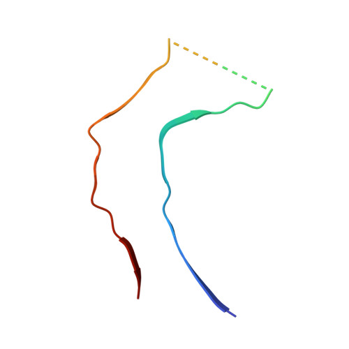
    Assembly of microtubule-associated protein tau into filamentous inclusions underlies a range of neurodegenerative diseases. Tau filaments adopt different conformations in Alzheimer's and Pick's diseases. Here, we used cryo- and immuno- electron microscopy to characterise filaments that were assembled from recombinant full-length human tau with four (2N4R) or three (2N3R) microtubule-binding repeats in the presence of heparin. 2N4R tau assembles into multiple types of filaments, and the structures of three types reveal similar 'kinked hairpin' folds, in which the second and third repeats pack against each other. 2N3R tau filaments are structurally homogeneous, and adopt a dimeric core, where the third repeats of two tau molecules pack in a parallel manner. The heparin-induced tau filaments differ from those of Alzheimer's or Pick's disease, which have larger cores with different repeat compositions. Our results illustrate the structural versatility of amyloid filaments, and raise questions about the relevance of in vitro assembly.


  • Organizational Affiliation
    • MRC Laboratory of Molecular Biology, Cambridge, United Kingdom.

Macromolecule Content 

  • Total Structure Weight: 15.57 kDa 
  • Atom Count: 846 
  • Modeled Residue Count: 111 
  • Deposited Residue Count: 144 
  • Unique protein chains: 1

Macromolecules

Find similar proteins by:|  3D Structure
Entity ID: 1
MoleculeChains  Sequence LengthOrganismDetailsImage
Microtubule-associated protein tau
A, B, C
48Homo sapiensMutation(s): 0 
Gene Names: MAPTMAPTLMTBT1TAU
UniProt & NIH Common Fund Data Resources
Find proteins for P10636 (Homo sapiens)
Explore P10636 
Go to UniProtKB:  P10636
PHAROS:  P10636
GTEx:  ENSG00000186868 
Entity Groups
Sequence Clusters30% Identity50% Identity70% Identity90% Identity95% Identity100% Identity
UniProt GroupP10636
Sequence Annotations
Expand
Reference Sequence

Experimental Data & Validation

Experimental Data

  • Method: ELECTRON MICROSCOPY
  • Resolution: 3.30 Å
  • Aggregation State: FILAMENT 
  • Reconstruction Method: SINGLE PARTICLE 
EM Software:
TaskSoftware PackageVersion
RECONSTRUCTIONRELION3.0
MODEL REFINEMENTREFMAC5.8.0236
MODEL REFINEMENTPHENIXdev-2919-000

Structure Validation

View Full Validation Report


Entry History 

& Funding Information

Deposition Data


Funding OrganizationLocationGrant Number
Medical Research Council (United Kingdom)United KingdomMC_UP_A025_1013
Medical Research Council (United Kingdom)United KingdomMC_U105184291

Revision History  (Full details and data files)

  • Version 1.0: 2019-02-27
    Type: Initial release
  • Version 1.1: 2024-05-15
    Changes: Data collection, Database references