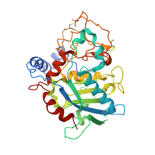
Structure-based mechanism of cysteine-switch latency and of catalysis by pappalysin-family metallopeptidases.
Guevara, T., Rodriguez-Banqueri, A., Ksiazek, M., Potempa, J., Gomis-Ruth, F.X.(2020) IUCrJ 7: 18-29
- PubMed: 31949901
- DOI: https://doi.org/10.1107/S2052252519013848
- Primary Citation of Related Structures:
6R7U, 6R7V, 6R7W - PubMed Abstract:
Tannerella forsythia is an oral dysbiotic periodontopathogen involved in severe human periodontal disease. As part of its virulence factor armamentarium, at the site of colonization it secretes mirolysin, a metallopeptidase of the unicellular pappalysin family, as a zymogen that is proteolytically auto-activated extracellularly at the Ser54-Arg55 bond. Crystal structures of the catalytically impaired promirolysin point mutant E225A at 1.4 and 1.6 Å revealed that latency is exerted by an N-terminal 34-residue pro-segment that shields the front surface of the 274-residue catalytic domain, thus preventing substrate access. The catalytic domain conforms to the metzincin clan of metallopeptidases and contains a double calcium site, which acts as a calcium switch for activity. The pro-segment traverses the active-site cleft in the opposite direction to the substrate, which precludes its cleavage. It is anchored to the mature enzyme through residue Arg21, which intrudes into the specificity pocket in cleft sub-site S 1 '. Moreover, residue Cys23 within a conserved cysteine-glycine motif blocks the catalytic zinc ion by a cysteine-switch mechanism, first described for mammalian matrix metallopeptidases. In addition, a 1.5 Å structure was obtained for a complex of mature mirolysin and a tetradecapeptide, which filled the cleft from sub-site S 1 ' to S 6 '. A citrate molecule in S 1 completed a product-complex mimic that unveiled the mechanism of substrate binding and cleavage by mirolysin, the catalytic domain of which was already preformed in the zymogen. These results, including a preference for cleavage before basic residues, are likely to be valid for other unicellular pappalysins derived from archaea, bacteria, cyanobacteria, algae and fungi, including archetypal ulilysin from Methanosarcina acetivorans . They may further apply, at least in part, to the multi-domain orthologues of higher organisms.
- Proteolysis Laboratory, Department of Structural Biology, Molecular Biology Institute of Barcelona, CSIC, Barcelona Science Park, Helix Building, c/ Baldiri Reixac, 15-21, 08028 Barcelona, Catalonia, Spain.
Organizational Affiliation:


