6RPG

Structure of human Programmed cell death 1 ligand 1 (PD-L1) with inhibitor


Experimental Data Snapshot

  • Method: X-RAY DIFFRACTION
  • Resolution: 2.70 Å
  • R-Value Free: 0.281 
  • R-Value Work: 0.248 
  • R-Value Observed: 0.251 

wwPDB Validation   3D Report Full Report


Ligand Structure Quality Assessment 


This is version 1.3 of the entry. See complete history


Literature

Design, Synthesis, Evaluation, and Structural Studies ofC2-Symmetric Small Molecule Inhibitors of Programmed Cell Death-1/Programmed Death-Ligand 1 Protein-Protein Interaction.

Basu, S.Yang, J.Xu, B.Magiera-Mularz, K.Skalniak, L.Musielak, B.Kholodovych, V.Holak, T.A.Hu, L.

(2019) J Med Chem 62: 7250-7263

  • DOI: https://doi.org/10.1021/acs.jmedchem.9b00795
  • Primary Citation of Related Structures:  
    6RPG

  • PubMed Abstract: 

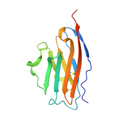
    A series of C 2 -symmetric inhibitors was designed and evaluated for inhibitory activity against the programmed cell death-1/programmed death-ligand 1(PD-1/PD-L1) protein-protein interaction (PPI) in a homogenous time-resolved fluorescence (HTRF) assay and PD-1 signaling in cell-based coculture assays. C 2 -symmetric inhibitors 2a (LH1306) and 2b (LH1307) exhibited IC 50 values of 25 and 3.0 nM, respectively, in the HTRF assay. While 2a was ∼3.8-fold more potent than previously reported inhibitor 1a , 2b could not be differentiated from 1b due to their high potency and the limit of our HTRF assay conditions. In one cell-based coculture PD-1 signaling assay, 2a and 2b were 8.2- and 2.8-fold more potent in inhibiting PD-1 signaling than 1a and 1b , respectively. NMR and X-ray cocrystal structural studies provided more structural insights into the interaction between 2b and PD-L1; 2b binds to PD-L1 at the PD-1 binding site and induces the formation of a more symmetrically arranged PD-L1 homodimer than that previously reported for other inhibitors.


  • Organizational Affiliation

    Department of Organic Chemistry, Faculty of Chemistry , Jagiellonian University , Gronostajowa 2 , 30-387 Krakow , Poland.


Macromolecules
Find similar proteins by:  (by identity cutoff)  |  3D Structure
Entity ID: 1
MoleculeChains Sequence LengthOrganismDetailsImage
Programmed cell death 1 ligand 1
A, B
128Homo sapiensMutation(s): 0 
Gene Names: CD274B7H1PDCD1L1PDCD1LG1PDL1
UniProt & NIH Common Fund Data Resources
Find proteins for Q9NZQ7 (Homo sapiens)
Explore Q9NZQ7 
Go to UniProtKB:  Q9NZQ7
PHAROS:  Q9NZQ7
GTEx:  ENSG00000120217 
Entity Groups  
Sequence Clusters30% Identity50% Identity70% Identity90% Identity95% Identity100% Identity
UniProt GroupQ9NZQ7
Sequence Annotations
Expand
  • Reference Sequence
Small Molecules
Ligands 1 Unique
IDChains Name / Formula / InChI Key2D Diagram3D Interactions
KDW
Query on KDW

Download Ideal Coordinates CCD File 
C [auth B]~{N}-[2-[[4-[[3-[3-[[4-[(2-acetamidoethylamino)methyl]-5-[(5-cyanopyridin-3-yl)methoxy]-2-methyl-phenoxy]methyl]-2-methyl-phenyl]-2-methyl-phenyl]methoxy]-2-[(5-cyanopyridin-3-yl)methoxy]-5-methyl-phenyl]methylamino]ethyl]ethanamide
C54 H58 N8 O6
RNCINFJGGSQEHN-UHFFFAOYSA-N
Experimental Data & Validation

Experimental Data

  • Method: X-RAY DIFFRACTION
  • Resolution: 2.70 Å
  • R-Value Free: 0.281 
  • R-Value Work: 0.248 
  • R-Value Observed: 0.251 
  • Space Group: P 31 2 1
Unit Cell:
Length ( Å )Angle ( ˚ )
a = 75.894α = 90
b = 75.894β = 90
c = 98.205γ = 120
Software Package:
Software NamePurpose
PHENIXrefinement
XDSdata reduction
SCALAdata scaling
PHASERphasing

Structure Validation

View Full Validation Report


Ligand Structure Quality Assessment 


Entry History & Funding Information

Deposition Data


Funding OrganizationLocationGrant Number
European UnionPolandPOIR.04.04.00-00-420F/17-00

Revision History  (Full details and data files)

  • Version 1.0: 2019-07-24
    Type: Initial release
  • Version 1.1: 2019-08-07
    Changes: Data collection, Database references
  • Version 1.2: 2019-08-21
    Changes: Data collection, Database references
  • Version 1.3: 2024-01-24
    Changes: Data collection, Database references, Refinement description