6RQ1

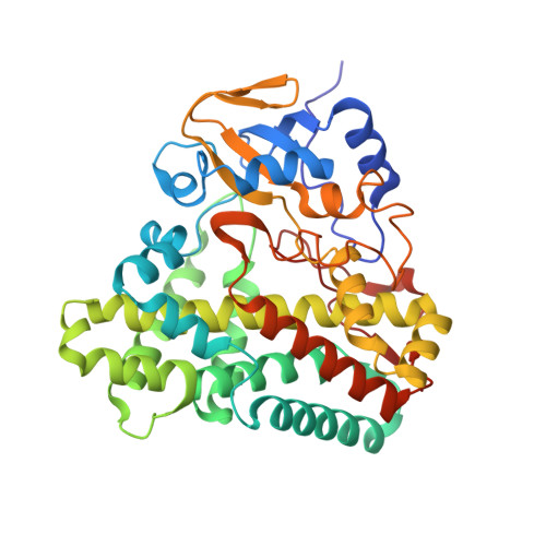
CYP121 in complex with 2-methyl dicyclotyrosine


Experimental Data Snapshot

  • Method: X-RAY DIFFRACTION
  • Resolution: 1.49 Å
  • R-Value Free: 0.196 
  • R-Value Work: 0.172 
  • R-Value Observed: 0.173 

Starting Model: experimental
View more details

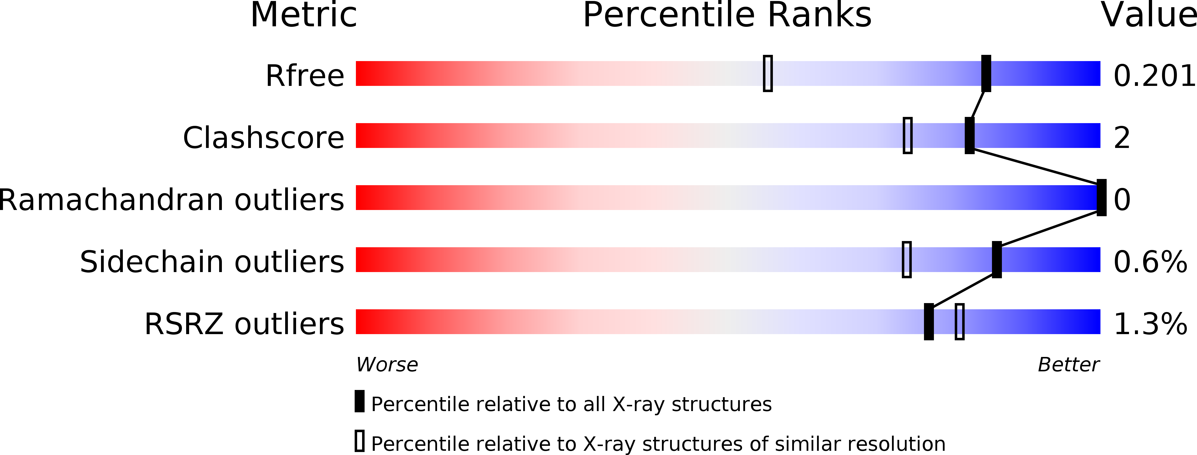
wwPDB Validation   3D Report Full Report


Ligand Structure Quality Assessment 


This is version 1.2 of the entry. See complete history


Literature

Structure-Activity Relationships of cyclo (l-Tyrosyl-l-tyrosine) Derivatives Binding to Mycobacterium tuberculosis CYP121: Iodinated Analogues Promote Shift to High-Spin Adduct.

Rajput, S.McLean, K.J.Poddar, H.Selvam, I.R.Nagalingam, G.Triccas, J.A.Levy, C.W.Munro, A.W.Hutton, C.A.

(2019) J Med Chem 62: 9792-9805

  • DOI: https://doi.org/10.1021/acs.jmedchem.9b01199
  • Primary Citation of Related Structures:  
    6RQ0, 6RQ1, 6RQ3, 6RQ5, 6RQ6, 6RQ8, 6RQ9, 6RQB, 6RQD, 6RQE

  • PubMed Abstract: 

    A series of analogues of cyclo (l-tyrosyl-l-tyrosine), the substrate of the Mycobacterium tuberculosis enzyme CYP121, have been synthesized and analyzed by UV-vis and electron paramagnetic resonance spectroscopy and by X-ray crystallography. The introduction of iodine substituents onto cyclo (l-tyrosyl-l-tyrosine) results in sub-μM binding affinity for the CYP121 enzyme and a complete shift to the high-spin state of the heme Fe III . The introduction of halogens that are able to interact with heme groups is thus a feasible approach to the development of next-generation, tight binding inhibitors of the CYP121 enzyme, in the search for novel antitubercular compounds.


  • Organizational Affiliation

    School of Chemistry and Bio21 Molecular Science and Biotechnology Institute , University of Melbourne , 30 Flemington Road , Parkville , Victoria 3010 , Australia.


Macromolecules
Find similar proteins by:  (by identity cutoff)  |  3D Structure
Entity ID: 1
MoleculeChains Sequence LengthOrganismDetailsImage
Mycocyclosin synthase396Mycobacterium tuberculosisMutation(s): 0 
Gene Names: cyp121MT2336
EC: 1.14.19.70
UniProt
Find proteins for P9WPP7 (Mycobacterium tuberculosis (strain ATCC 25618 / H37Rv))
Explore P9WPP7 
Go to UniProtKB:  P9WPP7
Entity Groups  
Sequence Clusters30% Identity50% Identity70% Identity90% Identity95% Identity100% Identity
UniProt GroupP9WPP7
Sequence Annotations
Expand
  • Reference Sequence
Small Molecules
Ligands 4 Unique
IDChains Name / Formula / InChI Key2D Diagram3D Interactions
HEM
Query on HEM

Download Ideal Coordinates CCD File 
C [auth A]PROTOPORPHYRIN IX CONTAINING FE
C34 H32 Fe N4 O4
KABFMIBPWCXCRK-RGGAHWMASA-L
KE5 (Subject of Investigation/LOI)
Query on KE5

Download Ideal Coordinates CCD File 
D [auth A](3~{S},6~{S})-3-[(4-hydroxyphenyl)methyl]-6-[(2-methyl-4-oxidanyl-phenyl)methyl]piperazine-2,5-dione
C19 H20 N2 O4
OLLRPTQZHVLIEA-IRXDYDNUSA-N
MES
Query on MES

Download Ideal Coordinates CCD File 
B [auth A]2-(N-MORPHOLINO)-ETHANESULFONIC ACID
C6 H13 N O4 S
SXGZJKUKBWWHRA-UHFFFAOYSA-N
SO4
Query on SO4

Download Ideal Coordinates CCD File 
E [auth A],
F [auth A],
G [auth A],
H [auth A]
SULFATE ION
O4 S
QAOWNCQODCNURD-UHFFFAOYSA-L
Experimental Data & Validation

Experimental Data

  • Method: X-RAY DIFFRACTION
  • Resolution: 1.49 Å
  • R-Value Free: 0.196 
  • R-Value Work: 0.172 
  • R-Value Observed: 0.173 
  • Space Group: P 65 2 2
Unit Cell:
Length ( Å )Angle ( ˚ )
a = 77.8α = 90
b = 77.8β = 90
c = 263.72γ = 120
Software Package:
Software NamePurpose
PHENIXrefinement
PDB_EXTRACTdata extraction
xia2data reduction
DIALSdata scaling
PHASERphasing

Structure Validation

View Full Validation Report


Ligand Structure Quality Assessment 


Entry History & Funding Information

Deposition Data


Funding OrganizationLocationGrant Number
Biotechnology and Biological Sciences Research CouncilUnited Kingdom--

Revision History  (Full details and data files)

  • Version 1.0: 2020-04-22
    Type: Initial release
  • Version 1.1: 2020-11-18
    Changes: Database references
  • Version 1.2: 2024-01-24
    Changes: Data collection, Database references, Refinement description