6SIU

Crystal structure of IbpAFic2 covalently tethered to Cdc42


Experimental Data Snapshot

  • Method: X-RAY DIFFRACTION
  • Resolution: 2.49 Å
  • R-Value Free: 0.239 
  • R-Value Work: 0.190 
  • R-Value Observed: 0.193 

wwPDB Validation   3D Report Full Report


Ligand Structure Quality Assessment 


This is version 1.3 of the entry. See complete history


Literature

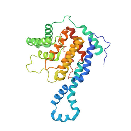
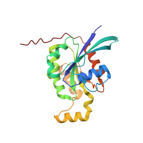
Identification of targets of AMPylating Fic enzymes by co-substrate-mediated covalent capture.

Gulen, B.Rosselin, M.Fauser, J.Albers, M.F.Pett, C.Krisp, C.Pogenberg, V.Schluter, H.Hedberg, C.Itzen, A.

(2020) Nat Chem 12: 732-739

  • DOI: https://doi.org/10.1038/s41557-020-0484-6
  • Primary Citation of Related Structures:  
    6SIU

  • PubMed Abstract: 

    Various pathogenic bacteria use post-translational modifications to manipulate the central components of host cell functions. Many of the enzymes released by these bacteria belong to the large Fic family, which modify targets with nucleotide monophosphates. The lack of a generic method for identifying the cellular targets of Fic family enzymes hinders investigation of their role and the effect of the post-translational modification. Here, we establish an approach that uses reactive co-substrate-linked enzymes for proteome profiling. We combine synthetic thiol-reactive nucleotide derivatives with recombinantly produced Fic enzymes containing strategically placed cysteines in their active sites to yield reactive binary probes for covalent substrate capture. The binary complexes capture their targets from cell lysates and permit subsequent identification. Furthermore, we determined the structures of low-affinity ternary enzyme-nucleotide-substrate complexes by applying a covalent-linking strategy. This approach thus allows target identification of the Fic enzymes from both bacteria and eukarya.


  • Organizational Affiliation

    Center for Integrated Protein Science Munich (CIPSM), Department of Chemistry, Technical University of Munich, Garching, Germany.


Macromolecules
Find similar proteins by:  (by identity cutoff)  |  3D Structure
Entity ID: 1
MoleculeChains Sequence LengthOrganismDetailsImage
Protein adenylyltransferase and cysteine protease IbpA
A, B
316Histophilus somni 2336Mutation(s): 1 
Gene Names: ibpAp76HSM_1489
EC: 2.7.7 (PDB Primary Data), 3.4.22 (PDB Primary Data)
UniProt
Find proteins for Q06277 (Histophilus somni (strain 2336))
Explore Q06277 
Go to UniProtKB:  Q06277
Entity Groups  
Sequence Clusters30% Identity50% Identity70% Identity90% Identity95% Identity100% Identity
UniProt GroupQ06277
Sequence Annotations
Expand
  • Reference Sequence
Find similar proteins by:  (by identity cutoff)  |  3D Structure
Entity ID: 2
MoleculeChains Sequence LengthOrganismDetailsImage
Cell division control protein 42 homolog
C, D
192Homo sapiensMutation(s): 0 
Gene Names: CDC42
EC: 3.6.5.2
UniProt & NIH Common Fund Data Resources
Find proteins for P60953 (Homo sapiens)
Explore P60953 
Go to UniProtKB:  P60953
PHAROS:  P60953
GTEx:  ENSG00000070831 
Entity Groups  
Sequence Clusters30% Identity50% Identity70% Identity90% Identity95% Identity100% Identity
UniProt GroupP60953
Sequence Annotations
Expand
  • Reference Sequence
Small Molecules
Ligands 4 Unique
IDChains Name / Formula / InChI Key2D Diagram3D Interactions
GDP
Query on GDP

Download Ideal Coordinates CCD File 
M [auth C],
Q [auth D]
GUANOSINE-5'-DIPHOSPHATE
C10 H15 N5 O11 P2
QGWNDRXFNXRZMB-UUOKFMHZSA-N
LJN
Query on LJN

Download Ideal Coordinates CCD File 
O [auth C],
S [auth D]
[(2~{R},3~{S},4~{R},5~{R})-5-[4-(acetamidomethyl)-1,2,3-triazol-1-yl]-3,4-bis(oxidanyl)oxolan-2-yl]methyl dihydrogen phosphate
C10 H17 N4 O8 P
WKSDLIPBAPRDGN-ZYUZMQFOSA-N
SO4
Query on SO4

Download Ideal Coordinates CCD File 
E [auth A]
F [auth A]
G [auth A]
H [auth A]
I [auth B]
E [auth A],
F [auth A],
G [auth A],
H [auth A],
I [auth B],
J [auth B],
K [auth B],
L [auth B],
P [auth C],
T [auth D]
SULFATE ION
O4 S
QAOWNCQODCNURD-UHFFFAOYSA-L
GOL
Query on GOL

Download Ideal Coordinates CCD File 
N [auth C],
R [auth D]
GLYCEROL
C3 H8 O3
PEDCQBHIVMGVHV-UHFFFAOYSA-N
Experimental Data & Validation

Experimental Data

  • Method: X-RAY DIFFRACTION
  • Resolution: 2.49 Å
  • R-Value Free: 0.239 
  • R-Value Work: 0.190 
  • R-Value Observed: 0.193 
  • Space Group: P 21 21 21
Unit Cell:
Length ( Å )Angle ( ˚ )
a = 92.241α = 90
b = 102.213β = 90
c = 129.273γ = 90
Software Package:
Software NamePurpose
PHENIXrefinement
DIALSdata reduction
Aimlessdata scaling
STARANISOdata scaling
PHASERphasing

Structure Validation

View Full Validation Report


Ligand Structure Quality Assessment 


Entry History & Funding Information

Deposition Data


Funding OrganizationLocationGrant Number
Knut and Alice Wallenberg FoundationSweden--
German Research FoundationGermanySFB1035 B05
Alexander von Humboldt FoundationGermany3.1 - USA - 1187240 - HFST-P

Revision History  (Full details and data files)

  • Version 1.0: 2020-03-18
    Type: Initial release
  • Version 1.1: 2020-07-15
    Changes: Database references
  • Version 1.2: 2020-08-05
    Changes: Database references
  • Version 1.3: 2024-01-24
    Changes: Data collection, Database references, Refinement description