


Crystal Structures Combined with Molecular Dynamics Reveal Altered Flow of Water in the Active Site of W60C Chromobacterium violaceum omega-transaminase
Ruggieri, F., Gustafsson, C., Kimbung, R.Y., Walse, B., Logan, D.T., Berglund, P.Not Published
Experimental Data Snapshot
Starting Model: experimental
View more details
Not Published
Entity ID: 1 | |||||
|---|---|---|---|---|---|
| Molecule | Chains | Sequence Length | Organism | Details | Image |
| Aspartate aminotransferase family protein | 465 | Chromobacterium violaceum | Mutation(s): 1 Gene Names: bioK, CBW21_20070, NCTC8684_01926 EC: 2.6.1 | 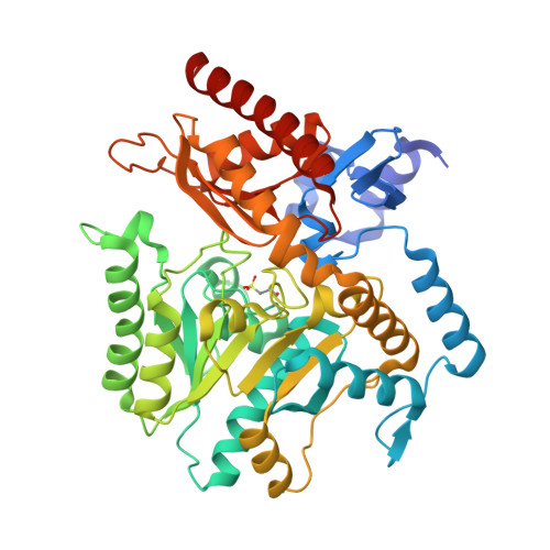 | |
UniProt | |||||
Find proteins for A0A1R0MXM9 (Chromobacterium violaceum) Explore A0A1R0MXM9 Go to UniProtKB: A0A1R0MXM9 | |||||
Entity Groups | |||||
| Sequence Clusters | 30% Identity50% Identity70% Identity90% Identity95% Identity100% Identity | ||||
| UniProt Group | A0A1R0MXM9 | ||||
Sequence AnnotationsExpand | |||||
Reference Sequence | |||||
| Ligands 2 Unique | |||||
|---|---|---|---|---|---|
| ID | Chains | Name / Formula / InChI Key | 2D Diagram | 3D Interactions | |
| PLP Download:Ideal Coordinates CCD File | F [auth A], J [auth B], K [auth C], L [auth D] | PYRIDOXAL-5'-PHOSPHATE C8 H10 N O6 P NGVDGCNFYWLIFO-UHFFFAOYSA-N |  | ||
| EDO Download:Ideal Coordinates CCD File | E [auth A], G [auth B], H [auth B], I [auth B] | 1,2-ETHANEDIOL C2 H6 O2 LYCAIKOWRPUZTN-UHFFFAOYSA-N |  | ||
| Modified Residues 1 Unique | |||||
|---|---|---|---|---|---|
| ID | Chains | Type | Formula | 2D Diagram | Parent |
| OCS Query on OCS | A, B, C, D | L-PEPTIDE LINKING | C3 H7 N O5 S |  | CYS |
| Length ( Å ) | Angle ( ˚ ) |
|---|---|
| a = 61.221 | α = 75.03 |
| b = 62.196 | β = 81.31 |
| c = 118.423 | γ = 75.3 |
| Software Name | Purpose |
|---|---|
| REFMAC | refinement |
| XDS | data reduction |
| Aimless | data scaling |
| PHASER | phasing |
| PDB_EXTRACT | data extraction |
| Funding Organization | Location | Grant Number |
|---|---|---|
| European Union | Sweden | 634200 |


