6ST3 | pdb_00006st3

HIF prolyl hydroxylase 2 (PHD2/ EGLN1) in complex with 4-hydroxy-N-(4-phenoxybenzyl)-2-(1H-pyrazol-1-yl)pyrimidine-5-carboxamide


Experimental Data Snapshot

  • Method: X-RAY DIFFRACTION
  • Resolution: 2.43 Å
  • R-Value Free: 
    0.217 (Depositor), 0.221 (DCC) 
  • R-Value Work: 
    0.192 (Depositor), 0.194 (DCC) 
  • R-Value Observed: 
    0.194 (Depositor) 

Starting Model: experimental
View more details

wwPDB Validation 3D Report Full Report

Validation slider image for 6ST3

Ligand Structure Quality Assessment 


This is version 1.2 of the entry. See complete history

Literature

Structure-Activity Relationship and Crystallographic Studies on 4-Hydroxypyrimidine HIF Prolyl Hydroxylase Domain Inhibitors.

Holt-Martyn, J.P.Chowdhury, R.Tumber, A.Yeh, T.L.Abboud, M.I.Lippl, K.Lohans, C.T.Langley, G.W.Figg Jr., W.McDonough, M.A.Pugh, C.W.Ratcliffe, P.J.Schofield, C.J.

(2020) ChemMedChem 15: 270-273

  • DOI: https://doi.org/10.1002/cmdc.201900557
  • Primary Citation Related Structures: 
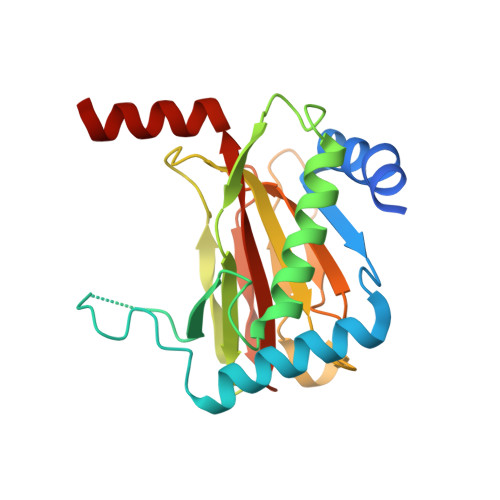
    6ST3

  • PubMed Abstract: 

    The 2-oxoglutarate-dependent hypoxia inducible factor prolyl hydroxylases (PHDs) are targets for treatment of a variety of diseases including anaemia. One PHD inhibitor is approved for use for the treatment of renal anaemia and others are in late stage clinical trials. The number of reported templates for PHD inhibition is limited. We report structure-activity relationship and crystallographic studies on a promising class of 4-hydroxypyrimidine-containing PHD inhibitors.


  • Organizational Affiliation
    • Department of Chemistry, University of Oxford Chemistry Research Laboratory, 12 Mansfield Road, Oxford, OX1 3TA, UK.

Macromolecule Content 

  • Total Structure Weight: 53.05 kDa 
  • Atom Count: 3,397 
  • Modeled Residue Count: 419 
  • Deposited Residue Count: 466 
  • Unique protein chains: 1

Macromolecules

Find similar proteins by:|  3D Structure
Entity ID: 1
MoleculeChains  Sequence LengthOrganismDetailsImage
Egl nine homolog 1
A, B
233Homo sapiensMutation(s): 0 
Gene Names: EGLN1C1orf12PNAS-118PNAS-137
EC: 1.14.11.29
UniProt & NIH Common Fund Data Resources
Find proteins for Q9GZT9 (Homo sapiens)
Explore Q9GZT9 
Go to UniProtKB:  Q9GZT9
PHAROS:  Q9GZT9
GTEx:  ENSG00000135766 
Entity Groups
Sequence Clusters30% Identity50% Identity70% Identity90% Identity95% Identity100% Identity
UniProt GroupQ9GZT9
Sequence Annotations
Expand
Reference Sequence

Small Molecules

Ligands 3 Unique
IDChains Name / Formula / InChI Key2D Diagram3D Interactions
LUW
(Subject of Investigation/LOI)

Query on LUW


Download:Ideal Coordinates CCD File
C [auth A],
G [auth B]
4-oxidanyl-~{N}-[(4-phenoxyphenyl)methyl]-2-pyrazol-1-yl-pyrimidine-5-carboxamide
C21 H17 N5 O3
WBANHLSPTRNPCR-UHFFFAOYSA-N
MN

Query on MN


Download:Ideal Coordinates CCD File
E [auth A],
F [auth B]
MANGANESE (II) ION
Mn
WAEMQWOKJMHJLA-UHFFFAOYSA-N
FMT

Query on FMT


Download:Ideal Coordinates CCD File
D [auth A],
H [auth B]
FORMIC ACID
C H2 O2
BDAGIHXWWSANSR-UHFFFAOYSA-N

Experimental Data & Validation

Experimental Data

  • Method: X-RAY DIFFRACTION
  • Resolution: 2.43 Å
  • R-Value Free:  0.217 (Depositor), 0.221 (DCC) 
  • R-Value Work:  0.192 (Depositor), 0.194 (DCC) 
  • R-Value Observed: 0.194 (Depositor) 
Space Group: P 21 2 21
Unit Cell:
Length ( Å )Angle ( ˚ )
a = 72.87α = 90
b = 80.58β = 90
c = 83.92γ = 90
Software Package:
Software NamePurpose
Aimlessdata scaling
PHENIXrefinement
PDB_EXTRACTdata extraction
MOSFLMdata reduction
PHASERphasing

Structure Validation

View Full Validation Report


Ligand Structure Quality Assessment 


Entry History 

Deposition Data

Revision History  (Full details and data files)

  • Version 1.0: 2019-12-04
    Type: Initial release
  • Version 1.1: 2020-02-19
    Changes: Database references
  • Version 1.2: 2024-01-24
    Changes: Data collection, Database references, Derived calculations, Refinement description