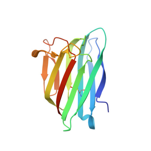
6SVK

human Myeloid-derived growth factor (MYDGF)


Experimental Data Snapshot

  • Method: X-RAY DIFFRACTION
  • Resolution: 1.60 Å
  • R-Value Free: 0.247 
  • R-Value Work: 0.218 
  • R-Value Observed: 0.219 

wwPDB Validation   3D Report Full Report


This is version 1.2 of the entry. See complete history


Literature

Crystal structure and receptor-interacting residues of MYDGF - a protein mediating ischemic tissue repair.

Ebenhoch, R.Akhdar, A.Reboll, M.R.Korf-Klingebiel, M.Gupta, P.Armstrong, J.Huang, Y.Frego, L.Rybina, I.Miglietta, J.Pekcec, A.Wollert, K.C.Nar, H.

(2019) Nat Commun 10: 5379-5379

  • DOI: https://doi.org/10.1038/s41467-019-13343-7
  • Primary Citation of Related Structures:  
    6SVK, 6SVL

  • PubMed Abstract: 

    Myeloid-derived growth factor (MYDGF) is a paracrine-acting protein that is produced by bone marrow-derived monocytes and macrophages to protect and repair the heart after myocardial infarction (MI). This effect can be used for the development of protein-based therapies for ischemic tissue repair, also beyond the sole application in heart tissue. Here, we report the X-ray structure of MYDGF and identify its functionally relevant receptor binding epitope. MYDGF consists of a 10-stranded β-sandwich with a folding topology showing no similarities to other cytokines or growth factors. By characterizing the epitope of a neutralizing antibody and utilizing functional assays to study the activity of surface patch-mutations, we were able to localize the receptor interaction interface to a region around two surface tyrosine residues 71 and 73 and an adjacent prominent loop structure of residues 97-101. These findings enable structure-guided protein engineering to develop modified MYDGF variants with potentially improved properties for clinical use.


  • Organizational Affiliation

    Boehringer Ingelheim Pharma GmbH & Co. KG, Birkendorfer Str. 65, 88397, Biberach an der Riss, Germany.


Macromolecules
Find similar proteins by:  (by identity cutoff)  |  3D Structure
Entity ID: 1
MoleculeChains Sequence LengthOrganismDetailsImage
Myeloid-derived growth factor
A, B
142Homo sapiensMutation(s): 0 
Gene Names: MYDGFC19orf10
UniProt & NIH Common Fund Data Resources
Find proteins for Q969H8 (Homo sapiens)
Explore Q969H8 
Go to UniProtKB:  Q969H8
PHAROS:  Q969H8
GTEx:  ENSG00000074842 
Entity Groups  
Sequence Clusters30% Identity50% Identity70% Identity90% Identity95% Identity100% Identity
UniProt GroupQ969H8
Sequence Annotations
Expand
  • Reference Sequence
Small Molecules
Ligands 1 Unique
IDChains Name / Formula / InChI Key2D Diagram3D Interactions
MLI
Query on MLI

Download Ideal Coordinates CCD File 
C [auth B]MALONATE ION
C3 H2 O4
OFOBLEOULBTSOW-UHFFFAOYSA-L
Experimental Data & Validation

Experimental Data

  • Method: X-RAY DIFFRACTION
  • Resolution: 1.60 Å
  • R-Value Free: 0.247 
  • R-Value Work: 0.218 
  • R-Value Observed: 0.219 
  • Space Group: P 31 2 1
Unit Cell:
Length ( Å )Angle ( ˚ )
a = 48.677α = 90
b = 48.677β = 90
c = 177.846γ = 120
Software Package:
Software NamePurpose
BUSTERrefinement
PDB_EXTRACTdata extraction
XDSdata reduction
Aimlessdata scaling
SHELXDphasing

Structure Validation

View Full Validation Report


Entry History 

Deposition Data

Revision History  (Full details and data files)

  • Version 1.0: 2019-11-27
    Type: Initial release
  • Version 1.1: 2019-12-04
    Changes: Database references
  • Version 1.2: 2024-11-13
    Changes: Data collection, Database references, Structure summary