6SZ5 | pdb_00006sz5

Human calmodulin bound to a peptide of human NADPH oxidase 5


Experimental Data Snapshot

  • Method: X-RAY DIFFRACTION
  • Resolution: 2.23 Å
  • R-Value Free: 
    0.294 (Depositor), 0.301 (DCC) 
  • R-Value Work: 
    0.219 (Depositor), 0.226 (DCC) 
  • R-Value Observed: 
    0.223 (Depositor) 

Starting Model: experimental
View more details

wwPDB Validation 3D Report Full Report

Validation slider image for 6SZ5

This is version 1.2 of the entry. See complete history

Literature

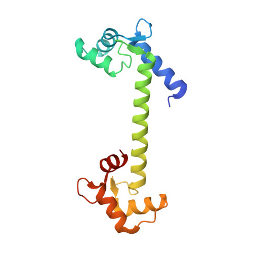
On the mechanism of calcium-dependent activation of NADPH oxidase 5 (NOX5).

Millana Fananas, E.Todesca, S.Sicorello, A.Masino, L.Pompach, P.Magnani, F.Pastore, A.Mattevi, A.

(2020) FEBS J 287: 2486-2503

  • DOI: https://doi.org/10.1111/febs.15160
  • Primary Citation Related Structures: 
    6SZ5

  • PubMed Abstract: 

    It is now accepted that reactive oxygen species (ROS) are not only dangerous oxidative agents but also chemical mediators of the redox cell signaling and innate immune response. A central role in ROS-controlled production is played by the NADPH oxidases (NOXs), a group of seven membrane-bound enzymes (NOX1-5 and DUOX1-2) whose unique function is to produce ROS. Here, we describe the regulation of NOX5, a widespread family member present in cyanobacteria, protists, plants, fungi, and the animal kingdom. We show that the calmodulin-like regulatory EF-domain of NOX5 is partially unfolded and detached from the rest of the protein in the absence of calcium. In the presence of calcium, the C-terminal lobe of the EF-domain acquires an ordered and more compact structure that enables its binding to the enzyme dehydrogenase (DH) domain. Our spectroscopic and mutagenesis studies further identified a set of conserved aspartate residues in the DH domain that are essential for NOX5 activation. Altogether, our work shows that calcium induces an unfolded-to-folded transition of the EF-domain that promotes direct interaction with a conserved regulatory region, resulting in NOX5 activation.


  • Organizational Affiliation
    • Department of Biology and Biotechnology "Lazzaro Spallanzani", University of Pavia, Italy.

Macromolecule Content 

  • Total Structure Weight: 181.25 kDa 
  • Atom Count: 1,362 
  • Modeled Residue Count: 165 
  • Deposited Residue Count: 1,587 
  • Unique protein chains: 2

Macromolecules

Find similar proteins by:|  3D Structure
Entity ID: 1
MoleculeChains  Sequence LengthOrganismDetailsImage
Calmodulin-2149Homo sapiensMutation(s): 0 
Gene Names: CALM2CAM2CAMB
UniProt & NIH Common Fund Data Resources
Find proteins for P0DP24 (Homo sapiens)
Explore P0DP24 
Go to UniProtKB:  P0DP24
PHAROS:  P0DP24
GTEx:  ENSG00000143933 
Entity Groups
Sequence Clusters30% Identity50% Identity70% Identity90% Identity95% Identity100% Identity
UniProt GroupP0DP24
Sequence Annotations
Expand
Reference Sequence
Find similar proteins by:|  3D Structure
Entity ID: 2
MoleculeChains  Sequence LengthOrganismDetailsImage
NADPH oxidase 5
B, C
719Homo sapiensMutation(s): 0 
EC: 1.6.3 (PDB Primary Data), 1 (UniProt)
UniProt & NIH Common Fund Data Resources
Find proteins for Q96PH1 (Homo sapiens)
Explore Q96PH1 
Go to UniProtKB:  Q96PH1
PHAROS:  Q96PH1
GTEx:  ENSG00000255346 
Entity Groups
Sequence Clusters30% Identity50% Identity70% Identity90% Identity95% Identity100% Identity
UniProt GroupQ96PH1
Sequence Annotations
Expand
Reference Sequence

Experimental Data & Validation

Experimental Data

  • Method: X-RAY DIFFRACTION
  • Resolution: 2.23 Å
  • R-Value Free:  0.294 (Depositor), 0.301 (DCC) 
  • R-Value Work:  0.219 (Depositor), 0.226 (DCC) 
  • R-Value Observed: 0.223 (Depositor) 
Space Group: P 21 21 21
Unit Cell:
Length ( Å )Angle ( ˚ )
a = 24.759α = 90
b = 60.84β = 90
c = 102.444γ = 90
Software Package:
Software NamePurpose
XDSdata reduction
Aimlessdata scaling
REFMACrefinement
PDB_EXTRACTdata extraction
PHASERphasing

Structure Validation

View Full Validation Report


Entry History 

& Funding Information

Deposition Data


Funding OrganizationLocationGrant Number
Italian Association for Cancer ResearchItalyIG19808

Revision History  (Full details and data files)

  • Version 1.0: 2019-12-11
    Type: Initial release
  • Version 1.1: 2020-07-01
    Changes: Database references
  • Version 1.2: 2024-01-24
    Changes: Data collection, Database references, Derived calculations, Refinement description