6TBE | pdb_00006tbe

LC3A in complex with (3R,4S,5R,6R)-5-hydroxy-6-((4-hydroxy-3-(4-hydroxy-3-isopentylbenzamido)-8-methyl-2-oxo-2H-chromen-7-yl)oxy)-3-methoxy-2,2-dimethyltetrahydro-2H-pyran-4-yl carbamate


Experimental Data Snapshot

  • Method: X-RAY DIFFRACTION
  • Resolution: 1.67 Å
  • R-Value Free: 
    0.212 (Depositor), 0.222 (DCC) 
  • R-Value Work: 
    0.186 (Depositor), 0.190 (DCC) 
  • R-Value Observed: 
    0.187 (Depositor) 

Starting Model: experimental
View more details

wwPDB Validation 3D Report Full Report

Validation slider image for 6TBE

Ligand Structure Quality Assessment 


This is version 1.2 of the entry. See complete history

Literature

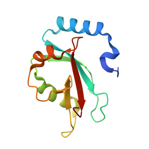
Demonstrating Ligandability of the LC3A and LC3B Adapter Interface.

Hartmann, M.Huber, J.Kramer, J.S.Heering, J.Pietsch, L.Stark, H.Odadzic, D.Bischoff, I.Furst, R.Schroder, M.Akutsu, M.Chaikuad, A.Dotsch, V.Knapp, S.Biondi, R.M.Rogov, V.V.Proschak, E.

(2021) J Med Chem 64: 3720-3746

  • DOI: https://doi.org/10.1021/acs.jmedchem.0c01564
  • Primary Citation Related Structures: 
    6TBE

  • PubMed Abstract: 

    Autophagy is the common name for a number of lysosome-based degradation pathways of cytosolic cargos. The key components of autophagy are members of Atg8 family proteins involved in almost all steps of the process, from autophagosome formation to their selective fusion with lysosomes. In this study, we show that the homologous members of the human Atg8 family proteins, LC3A and LC3B, are druggable by a small molecule inhibitor novobiocin. Structure-activity relationship (SAR) studies of the 4-hydroxy coumarin core scaffold were performed, supported by a crystal structure of the LC3A dihydronovobiocin complex. The study reports the first nonpeptide inhibitors for these protein interaction targets and will lay the foundation for the development of more potent chemical probes for the Atg8 protein family which may also find applications for the development of autophagy-mediated degraders (AUTACs).


  • Organizational Affiliation
    • Institute of Pharmaceutical Chemistry, Goethe-University Frankfurt, Max-von-Laue-Str. 9, 60438 Frankfurt, Germany.

Macromolecule Content 

  • Total Structure Weight: 14.77 kDa 
  • Atom Count: 1,142 
  • Modeled Residue Count: 116 
  • Deposited Residue Count: 119 
  • Unique protein chains: 1

Macromolecules

Find similar proteins by:|  3D Structure
Entity ID: 1
MoleculeChains  Sequence LengthOrganismDetailsImage
Microtubule-associated proteins 1A/1B light chain 3A119Homo sapiensMutation(s): 0 
Gene Names: MAP1LC3A
UniProt & NIH Common Fund Data Resources
Find proteins for Q9H492 (Homo sapiens)
Explore Q9H492 
Go to UniProtKB:  Q9H492
PHAROS:  Q9H492
GTEx:  ENSG00000101460 
Entity Groups
Sequence Clusters30% Identity50% Identity70% Identity90% Identity95% Identity100% Identity
UniProt GroupQ9H492
Sequence Annotations
Expand
Reference Sequence

Experimental Data & Validation

Experimental Data

  • Method: X-RAY DIFFRACTION
  • Resolution: 1.67 Å
  • R-Value Free:  0.212 (Depositor), 0.222 (DCC) 
  • R-Value Work:  0.186 (Depositor), 0.190 (DCC) 
  • R-Value Observed: 0.187 (Depositor) 
Space Group: I 41
Unit Cell:
Length ( Å )Angle ( ˚ )
a = 95.738α = 90
b = 95.738β = 90
c = 34.647γ = 90
Software Package:
Software NamePurpose
PHENIXrefinement
XDSdata reduction
XSCALEdata scaling
PHASERphasing

Structure Validation

View Full Validation Report


Ligand Structure Quality Assessment 


Entry History 

& Funding Information

Deposition Data


Funding OrganizationLocationGrant Number
German Federal Ministry for Education and ResearchGermanyDeutsches Konsortium fuer Translationale Krebsforschung
German Research FoundationGermanyPR 1405/4-1

Revision History  (Full details and data files)

  • Version 1.0: 2020-11-18
    Type: Initial release
  • Version 1.1: 2021-12-01
    Changes: Database references
  • Version 1.2: 2024-01-24
    Changes: Data collection, Refinement description