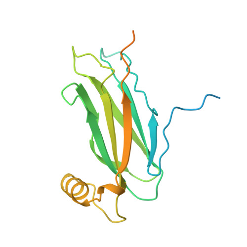
6TQS

The crystal structure of the MSP domain of human MOSPD2 in complex with the conventional FFAT motif of ORP1.


Experimental Data Snapshot

  • Method: X-RAY DIFFRACTION
  • Resolution: 2.25 Å
  • R-Value Free: 0.260 
  • R-Value Work: 0.217 
  • R-Value Observed: 0.219 

wwPDB Validation   3D Report Full Report


This is version 1.2 of the entry. See complete history


Literature

FFAT motif phosphorylation controls formation and lipid transfer function of inter-organelle contacts.

Di Mattia, T.Martinet, A.Ikhlef, S.McEwen, A.G.Nomine, Y.Wendling, C.Poussin-Courmontagne, P.Voilquin, L.Eberling, P.Ruffenach, F.Cavarelli, J.Slee, J.Levine, T.P.Drin, G.Tomasetto, C.Alpy, F.

(2020) EMBO J 39: e104369-e104369

  • DOI: https://doi.org/10.15252/embj.2019104369
  • Primary Citation of Related Structures:  
    6TQR, 6TQS, 6TQT, 6TQU

  • PubMed Abstract: 

    Organelles are physically connected in membrane contact sites. The endoplasmic reticulum possesses three major receptors, VAP-A, VAP-B, and MOSPD2, which interact with proteins at the surface of other organelles to build contacts. VAP-A, VAP-B, and MOSPD2 contain an MSP domain, which binds a motif named FFAT (two phenylalanines in an acidic tract). In this study, we identified a non-conventional FFAT motif where a conserved acidic residue is replaced by a serine/threonine. We show that phosphorylation of this serine/threonine is critical for non-conventional FFAT motifs (named Phospho-FFAT) to be recognized by the MSP domain. Moreover, structural analyses of the MSP domain alone or in complex with conventional and Phospho-FFAT peptides revealed new mechanisms of interaction. Based on these new insights, we produced a novel prediction algorithm, which expands the repertoire of candidate proteins with a Phospho-FFAT that are able to create membrane contact sites. Using a prototypical tethering complex made by STARD3 and VAP, we showed that phosphorylation is instrumental for the formation of ER-endosome contacts, and their sterol transfer function. This study reveals that phosphorylation acts as a general switch for inter-organelle contacts.


  • Organizational Affiliation

    Institut de Génétique et de Biologie Moléculaire et Cellulaire (IGBMC), Illkirch, France.


Macromolecules
Find similar proteins by:  (by identity cutoff)  |  3D Structure
Entity ID: 1
MoleculeChains Sequence LengthOrganismDetailsImage
Motile sperm domain-containing protein 2
A, B, C, D, E
A, B, C, D, E, F
216Homo sapiensMutation(s): 0 
Gene Names: MOSPD2
UniProt & NIH Common Fund Data Resources
Find proteins for Q8NHP6 (Homo sapiens)
Explore Q8NHP6 
Go to UniProtKB:  Q8NHP6
PHAROS:  Q8NHP6
GTEx:  ENSG00000130150 
Entity Groups  
Sequence Clusters30% Identity50% Identity70% Identity90% Identity95% Identity100% Identity
UniProt GroupQ8NHP6
Sequence Annotations
Expand
  • Reference Sequence

Find similar proteins by:  Sequence   |   3D Structure  

Entity ID: 2
MoleculeChains Sequence LengthOrganismDetailsImage
Oxysterol-binding protein-related protein 1
G, H, I, J, K
21Homo sapiensMutation(s): 0 
UniProt & NIH Common Fund Data Resources
Find proteins for Q9BXW6 (Homo sapiens)
Explore Q9BXW6 
Go to UniProtKB:  Q9BXW6
PHAROS:  Q9BXW6
GTEx:  ENSG00000141447 
Entity Groups  
Sequence Clusters30% Identity50% Identity70% Identity90% Identity95% Identity100% Identity
UniProt GroupQ9BXW6
Sequence Annotations
Expand
  • Reference Sequence
Small Molecules
Ligands 6 Unique
IDChains Name / Formula / InChI Key2D Diagram3D Interactions
1PE
Query on 1PE

Download Ideal Coordinates CCD File 
CA [auth F]PENTAETHYLENE GLYCOL
C10 H22 O6
JLFNLZLINWHATN-UHFFFAOYSA-N
TFA
Query on TFA

Download Ideal Coordinates CCD File 
BA [auth F]
N [auth A]
R [auth B]
U [auth C]
X [auth D]
BA [auth F],
N [auth A],
R [auth B],
U [auth C],
X [auth D],
Y [auth E]
trifluoroacetic acid
C2 H F3 O2
DTQVDTLACAAQTR-UHFFFAOYSA-N
PEG
Query on PEG

Download Ideal Coordinates CCD File 
V [auth C]DI(HYDROXYETHYL)ETHER
C4 H10 O3
MTHSVFCYNBDYFN-UHFFFAOYSA-N
SO4
Query on SO4

Download Ideal Coordinates CCD File 
AA [auth E],
DA [auth F],
P [auth A],
W [auth C],
Z [auth E]
SULFATE ION
O4 S
QAOWNCQODCNURD-UHFFFAOYSA-L
GOL
Query on GOL

Download Ideal Coordinates CCD File 
L [auth A],
M [auth A],
O [auth A],
S [auth B],
T [auth C]
GLYCEROL
C3 H8 O3
PEDCQBHIVMGVHV-UHFFFAOYSA-N
CL
Query on CL

Download Ideal Coordinates CCD File 
Q [auth A]CHLORIDE ION
Cl
VEXZGXHMUGYJMC-UHFFFAOYSA-M
Experimental Data & Validation

Experimental Data

  • Method: X-RAY DIFFRACTION
  • Resolution: 2.25 Å
  • R-Value Free: 0.260 
  • R-Value Work: 0.217 
  • R-Value Observed: 0.219 
  • Space Group: P 61 2 2
Unit Cell:
Length ( Å )Angle ( ˚ )
a = 126.874α = 90
b = 126.874β = 90
c = 184.449γ = 120
Software Package:
Software NamePurpose
XDSdata reduction
Aimlessdata scaling
PHASERphasing
PHENIXrefinement
PDB_EXTRACTdata extraction

Structure Validation

View Full Validation Report


Entry History & Funding Information

Deposition Data


Funding OrganizationLocationGrant Number
French National Research AgencyFranceANR-19-CE44-0003
French National Research AgencyFranceANR-10-INSB-05-01
French National Research AgencyFranceANR-10-LABX-0030-INRT
French National Research AgencyFranceANR-10-IDEX-0002-02.

Revision History  (Full details and data files)

  • Version 1.0: 2020-11-18
    Type: Initial release
  • Version 1.1: 2020-12-16
    Changes: Database references
  • Version 1.2: 2024-01-24
    Changes: Data collection, Database references, Refinement description