6TX3

HPF1 bound to catalytic fragment of PARP2

  • Classification: TRANSFERASE
  • Organism(s): Homo sapiens
  • Expression System: Escherichia coli BL21(DE3)
  • Mutation(s): Yes 

  • Deposited: 2020-01-13 Released: 2020-02-19 
  • Deposition Author(s): Suskiewicz, M.J., Ahel, I.
  • Funding Organization(s): Wellcome Trust, Biotechnology and Biological Sciences Research Council (BBSRC), Cancer Research UK, European Molecular Biology Organization (EMBO)

Experimental Data Snapshot

  • Method: X-RAY DIFFRACTION
  • Resolution: 2.96 Å
  • R-Value Free: 0.260 
  • R-Value Work: 0.229 
  • R-Value Observed: 0.231 

wwPDB Validation   3D Report Full Report


Ligand Structure Quality Assessment 


This is version 1.2 of the entry. See complete history


Literature

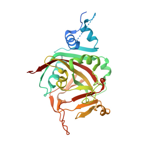
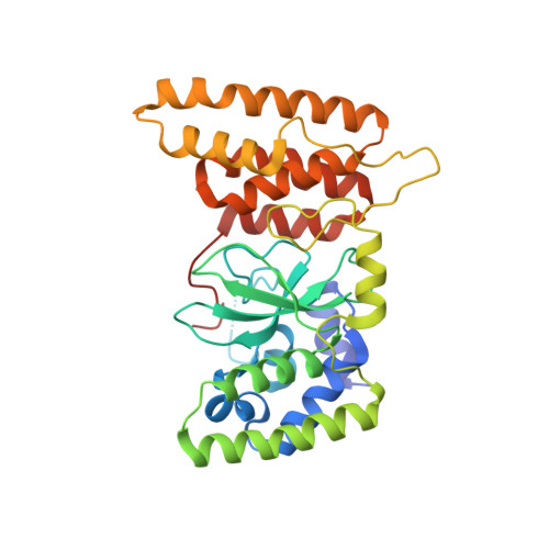
HPF1 completes the PARP active site for DNA damage-induced ADP-ribosylation.

Suskiewicz, M.J.Zobel, F.Ogden, T.E.H.Fontana, P.Ariza, A.Yang, J.C.Zhu, K.Bracken, L.Hawthorne, W.J.Ahel, D.Neuhaus, D.Ahel, I.

(2020) Nature 579: 598-602

  • DOI: https://doi.org/10.1038/s41586-020-2013-6
  • Primary Citation of Related Structures:  
    6TVH, 6TX1, 6TX2, 6TX3

  • PubMed Abstract: 

    The anti-cancer drug target poly(ADP-ribose) polymerase 1 (PARP1) and its close homologue, PARP2, are early responders to DNA damage in human cells 1,2 . After binding to genomic lesions, these enzymes use NAD + to modify numerous proteins with mono- and poly(ADP-ribose) signals that are important for the subsequent decompaction of chromatin and the recruitment of repair factors 3,4 . These post-translational modifications are predominantly serine-linked and require the accessory factor HPF1, which is specific for the DNA damage response and switches the amino acid specificity of PARP1 and PARP2 from aspartate or glutamate to serine residues 5-10 . Here we report a co-structure of HPF1 bound to the catalytic domain of PARP2 that, in combination with NMR and biochemical data, reveals a composite active site formed by residues from HPF1 and PARP1 or PARP2 . The assembly of this catalytic centre is essential for the addition of ADP-ribose moieties after DNA damage in human cells. In response to DNA damage and occupancy of the NAD + -binding site, the interaction of HPF1 with PARP1 or PARP2 is enhanced by allosteric networks that operate within the PARP proteins, providing an additional level of regulation in the induction of the DNA damage response. As HPF1 forms a joint active site with PARP1 or PARP2, our data implicate HPF1 as an important determinant of the response to clinical PARP inhibitors.


  • Organizational Affiliation

    Sir William Dunn School of Pathology, University of Oxford, Oxford, UK.


Macromolecules
Find similar proteins by:  (by identity cutoff)  |  3D Structure
Entity ID: 1
MoleculeChains Sequence LengthOrganismDetailsImage
Poly [ADP-ribose] polymerase 2,Poly [ADP-ribose] polymerase 2A [auth B]280Homo sapiensMutation(s): 0 
Gene Names: PARP2ADPRT2ADPRTL2
EC: 2.4.2.30 (PDB Primary Data), 2.4.2 (PDB Primary Data)
UniProt & NIH Common Fund Data Resources
Find proteins for Q9UGN5 (Homo sapiens)
Explore Q9UGN5 
Go to UniProtKB:  Q9UGN5
PHAROS:  Q9UGN5
GTEx:  ENSG00000129484 
Entity Groups  
Sequence Clusters30% Identity50% Identity70% Identity90% Identity95% Identity100% Identity
UniProt GroupQ9UGN5
Sequence Annotations
Expand
  • Reference Sequence
Find similar proteins by:  (by identity cutoff)  |  3D Structure
Entity ID: 2
MoleculeChains Sequence LengthOrganismDetailsImage
Histone PARylation factor 1B [auth A]318Homo sapiensMutation(s): 1 
Gene Names: HPF1C4orf27
UniProt & NIH Common Fund Data Resources
Find proteins for Q9NWY4 (Homo sapiens)
Explore Q9NWY4 
Go to UniProtKB:  Q9NWY4
PHAROS:  Q9NWY4
GTEx:  ENSG00000056050 
Entity Groups  
Sequence Clusters30% Identity50% Identity70% Identity90% Identity95% Identity100% Identity
UniProt GroupQ9NWY4
Sequence Annotations
Expand
  • Reference Sequence
Small Molecules
Ligands 1 Unique
IDChains Name / Formula / InChI Key2D Diagram3D Interactions
UHB (Subject of Investigation/LOI)
Query on UHB

Download Ideal Coordinates CCD File 
C [auth B]2-[4-[(2S,3S,4R,5R)-5-(6-aminopurin-9-yl)-3,4-bis(oxidanyl)oxolan-2-yl]carbonylpiperazin-1-yl]-N-(1-oxidanylidene-2,3-dihydroisoindol-4-yl)ethanamide
C24 H27 N9 O6
QEUOCRGVJJDDTK-VBFAUCSFSA-N
Experimental Data & Validation

Experimental Data

  • Method: X-RAY DIFFRACTION
  • Resolution: 2.96 Å
  • R-Value Free: 0.260 
  • R-Value Work: 0.229 
  • R-Value Observed: 0.231 
  • Space Group: P 61
Unit Cell:
Length ( Å )Angle ( ˚ )
a = 115.622α = 90
b = 115.622β = 90
c = 143.607γ = 120
Software Package:
Software NamePurpose
xia2data scaling
PHENIXrefinement
PDB_EXTRACTdata extraction
xia2data reduction
PHASERphasing

Structure Validation

View Full Validation Report


Ligand Structure Quality Assessment 


Entry History & Funding Information

Deposition Data


Funding OrganizationLocationGrant Number
Wellcome TrustUnited Kingdom101794
Wellcome TrustUnited Kingdom210634
Biotechnology and Biological Sciences Research Council (BBSRC)United KingdomB/R007195/1
Cancer Research UKUnited KingdomC35050/A22284
European Molecular Biology Organization (EMBO)European UnionALTF 879-2017

Revision History  (Full details and data files)

  • Version 1.0: 2020-02-19
    Type: Initial release
  • Version 1.1: 2020-04-08
    Changes: Database references
  • Version 1.2: 2024-05-01
    Changes: Data collection, Database references, Refinement description