6UKA | pdb_00006uka

Crystal structure of RHOG and ELMO complex


Experimental Data Snapshot

  • Method: X-RAY DIFFRACTION
  • Resolution: 2.40 Å
  • R-Value Free: 
    0.237 (Depositor), 0.225 (DCC) 
  • R-Value Work: 
    0.186 (Depositor) 
  • R-Value Observed: 
    0.191 (Depositor) 

Starting Model: experimental
View more details

wwPDB Validation 3D Report Full Report

Validation slider image for 6UKA

Ligand Structure Quality Assessment 


This is version 1.1 of the entry. See complete history

Literature

Structure of the DOCK2-ELMO1 complex provides insights into regulation of the auto-inhibited state.

Chang, L.Yang, J.Jo, C.H.Boland, A.Zhang, Z.McLaughlin, S.H.Abu-Thuraia, A.Killoran, R.C.Smith, M.J.Cote, J.F.Barford, D.

(2020) Nat Commun 11: 3464-3464

  • DOI: https://doi.org/10.1038/s41467-020-17271-9
  • Primary Citation Related Structures: 
    6TGB, 6TGC, 6UKA

  • PubMed Abstract: 

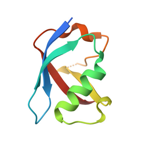
    DOCK (dedicator of cytokinesis) proteins are multidomain guanine nucleotide exchange factors (GEFs) for RHO GTPases that regulate intracellular actin dynamics. DOCK proteins share catalytic (DOCK DHR2 ) and membrane-associated (DOCK DHR1 ) domains. The structurally-related DOCK1 and DOCK2 GEFs are specific for RAC, and require ELMO (engulfment and cell motility) proteins for function. The N-terminal RAS-binding domain (RBD) of ELMO (ELMO RBD ) interacts with RHOG to modulate DOCK1/2 activity. Here, we determine the cryo-EM structures of DOCK2-ELMO1 alone, and as a ternary complex with RAC1, together with the crystal structure of a RHOG-ELMO2 RBD complex. The binary DOCK2-ELMO1 complex adopts a closed, auto-inhibited conformation. Relief of auto-inhibition to an active, open state, due to a conformational change of the ELMO1 subunit, exposes binding sites for RAC1 on DOCK2 DHR2 , and RHOG and BAI GPCRs on ELMO1. Our structure explains how up-stream effectors, including DOCK2 and ELMO1 phosphorylation, destabilise the auto-inhibited state to promote an active GEF.


  • Organizational Affiliation
    • MRC Laboratory of Molecular Biology, Cambridge, CB2 0QH, UK.

Macromolecule Content 

  • Total Structure Weight: 30.91 kDa 
  • Atom Count: 2,097 
  • Modeled Residue Count: 249 
  • Deposited Residue Count: 272 
  • Unique protein chains: 2

Macromolecules

Find similar proteins by:|  3D Structure
Entity ID: 1
MoleculeChains  Sequence LengthOrganismDetailsImage
Rho-related GTP-binding protein RhoG191Homo sapiensMutation(s): 0 
Gene Names: RHOGARHG
UniProt & NIH Common Fund Data Resources
Find proteins for P84095 (Homo sapiens)
Explore P84095 
Go to UniProtKB:  P84095
PHAROS:  P84095
GTEx:  ENSG00000177105 
Entity Groups
Sequence Clusters30% Identity50% Identity70% Identity90% Identity95% Identity100% Identity
UniProt GroupP84095
Sequence Annotations
Expand
Reference Sequence
Find similar proteins by:|  3D Structure
Entity ID: 2
MoleculeChains  Sequence LengthOrganismDetailsImage
Engulfment and cell motility protein 281Mus musculusMutation(s): 0 
Gene Names: Elmo2Kiaa1834
UniProt
Find proteins for Q8BHL5 (Mus musculus)
Explore Q8BHL5 
Go to UniProtKB:  Q8BHL5
Entity Groups
Sequence Clusters30% Identity50% Identity70% Identity90% Identity95% Identity100% Identity
UniProt GroupQ8BHL5
Sequence Annotations
Expand
Reference Sequence

Small Molecules

Experimental Data & Validation

Experimental Data

  • Method: X-RAY DIFFRACTION
  • Resolution: 2.40 Å
  • R-Value Free:  0.237 (Depositor), 0.225 (DCC) 
  • R-Value Work:  0.186 (Depositor) 
  • R-Value Observed: 0.191 (Depositor) 
Space Group: C 1 2 1
Unit Cell:
Length ( Å )Angle ( ˚ )
a = 100.279α = 90
b = 40.06β = 119.71
c = 73.93γ = 90
Software Package:
Software NamePurpose
PHENIXphasing
PHENIXrefinement
PROTEUM PLUSdata collection
PROTEUM PLUSdata scaling
PROTEUM PLUSdata reduction

Structure Validation

View Full Validation Report


Ligand Structure Quality Assessment 


Entry History 

& Funding Information

Deposition Data


Funding OrganizationLocationGrant Number
Canadian Institutes of Health Research (CIHR)Canada--

Revision History  (Full details and data files)

  • Version 1.0: 2020-08-12
    Type: Initial release
  • Version 1.1: 2023-10-11
    Changes: Data collection, Database references, Refinement description