6UX1 | pdb_00006ux1

Carbonic Anhydrase II Complexed with Salicylic Acid


Experimental Data Snapshot

  • Method: X-RAY DIFFRACTION
  • Resolution: 1.36 Å
  • R-Value Free: 
    0.167 (Depositor), 0.167 (DCC) 
  • R-Value Work: 
    0.154 (Depositor), 0.155 (DCC) 
  • R-Value Observed: 
    0.155 (Depositor) 

Starting Model: experimental
View more details

wwPDB Validation 3D Report Full Report

Validation slider image for 6UX1

Ligand Structure Quality Assessment 


This is version 1.1 of the entry. See complete history

Literature

Aspirin: A Suicide Inhibitor of Carbonic Anhydrase II.

Andring, J.Combs, J.McKenna, R.

(2020) Biomolecules 10

  • DOI: https://doi.org/10.3390/biom10040527
  • Primary Citation Related Structures: 
    6UX1

  • PubMed Abstract: 

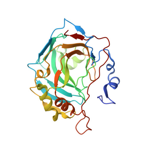
    Carbonic anhydrase II (CAII) is a metalloenzyme that catalyzes the reversible hydration/dehydration of CO 2 /HCO 3 - . In addition, CAII is attributed to other catalytic reactions, including esterase activity. Aspirin (acetyl-salicylic acid), an everyday over-the-counter drug, has both ester and carboxylic acid moieties. Recently, compounds with a carboxylic acid group have been shown to inhibit CAII. Hence, we hypothesized that Aspirin could act as a substrate for esterase activity, and the product salicylic acid (SA), an inhibitor of CAII. Here, we present the crystal structure of CAII in complex with SA, a product of CAII crystals pre-soaked with Aspirin, to 1.35Å resolution. In addition, we provide kinetic data to support the observation that CAII converts Aspirin to its deacetylated form, SA. This data may also explain the short half-life of Aspirin, with CAII so abundant in blood, and that Aspirin could act as a suicide inhibitor of CAII.


  • Organizational Affiliation
    • Department of Biochemistry and Molecular Biology, College of Medicine, University of Florida, Gainesville, FL 32610, USA.

Macromolecule Content 

  • Total Structure Weight: 30.12 kDa 
  • Atom Count: 2,351 
  • Modeled Residue Count: 258 
  • Deposited Residue Count: 260 
  • Unique protein chains: 1

Macromolecules

Find similar proteins by:|  3D Structure
Entity ID: 1
MoleculeChains  Sequence LengthOrganismDetailsImage
Carbonic anhydrase 2260Homo sapiensMutation(s): 0 
Gene Names: CA2
EC: 4.2.1.1 (PDB Primary Data), 4.2.1.69 (UniProt)
UniProt & NIH Common Fund Data Resources
Find proteins for P00918 (Homo sapiens)
Explore P00918 
Go to UniProtKB:  P00918
PHAROS:  P00918
GTEx:  ENSG00000104267 
Entity Groups
Sequence Clusters30% Identity50% Identity70% Identity90% Identity95% Identity100% Identity
UniProt GroupP00918
Sequence Annotations
Expand
Reference Sequence

Small Molecules

Ligands 4 Unique
IDChains Name / Formula / InChI Key2D Diagram3D Interactions
SAL
(Subject of Investigation/LOI)

Query on SAL


Download:Ideal Coordinates CCD File
D [auth A],
E [auth A],
F [auth A],
G [auth A]
2-HYDROXYBENZOIC ACID
C7 H6 O3
YGSDEFSMJLZEOE-UHFFFAOYSA-N
TRS

Query on TRS


Download:Ideal Coordinates CCD File
H [auth A]2-AMINO-2-HYDROXYMETHYL-PROPANE-1,3-DIOL
C4 H12 N O3
LENZDBCJOHFCAS-UHFFFAOYSA-O
GOL

Query on GOL


Download:Ideal Coordinates CCD File
C [auth A]GLYCEROL
C3 H8 O3
PEDCQBHIVMGVHV-UHFFFAOYSA-N
ZN

Query on ZN


Download:Ideal Coordinates CCD File
B [auth A]ZINC ION
Zn
PTFCDOFLOPIGGS-UHFFFAOYSA-N
Binding Affinity Annotations 
IDSourceBinding Affinity
SAL BindingDB:  6UX1 Ki: min: 7100, max: 7.40e+5 (nM) from 3 assay(s)
IC50: 6.80e+5 (nM) from 1 assay(s)

Experimental Data & Validation

Experimental Data

  • Method: X-RAY DIFFRACTION
  • Resolution: 1.36 Å
  • R-Value Free:  0.167 (Depositor), 0.167 (DCC) 
  • R-Value Work:  0.154 (Depositor), 0.155 (DCC) 
  • R-Value Observed: 0.155 (Depositor) 
Space Group: P 1 21 1
Unit Cell:
Length ( Å )Angle ( ˚ )
a = 42.427α = 90
b = 41.304β = 104.23
c = 72.028γ = 90
Software Package:
Software NamePurpose
PHENIXrefinement
Aimlessdata scaling
PDB_EXTRACTdata extraction
XDSdata reduction
PHENIXphasing

Structure Validation

View Full Validation Report


Ligand Structure Quality Assessment 


Entry History 

Deposition Data

Revision History  (Full details and data files)

  • Version 1.0: 2020-04-22
    Type: Initial release
  • Version 1.1: 2023-10-11
    Changes: Data collection, Database references, Derived calculations, Refinement description