6V9C | pdb_00006v9c

Crystal structure of FGFR4 kinase domain in complex with covalent inhibitor


Experimental Data Snapshot

  • Method: X-RAY DIFFRACTION
  • Resolution: 1.90 Å
  • R-Value Free: 
    0.227 (Depositor), 0.230 (DCC) 
  • R-Value Work: 
    0.199 (Depositor), 0.199 (DCC) 
  • R-Value Observed: 
    0.201 (Depositor) 

Starting Model: experimental
View more details

wwPDB Validation 3D Report Full Report

Validation slider image for 6V9C

Ligand Structure Quality Assessment 


This is version 1.3 of the entry. See complete history

Literature

Discovery of Selective, Covalent FGFR4 Inhibitors with Antitumor Activity in Models of Hepatocellular Carcinoma.

Liu, H.Niu, D.Tham Sjin, R.T.Dubrovskiy, A.Zhu, Z.McDonald, J.J.Fahnoe, K.Wang, Z.Munson, M.Scholte, A.Barrague, M.Fitzgerald, M.Liu, J.Kothe, M.Sun, F.Murtie, J.Ge, J.Rocnik, J.Harvey, D.Ospina, B.Perron, K.Zheng, G.Shehu, E.D'Agostino, L.A.

(2020) ACS Med Chem Lett 11: 1899-1904

  • DOI: https://doi.org/10.1021/acsmedchemlett.9b00601
  • Primary Citation Related Structures: 
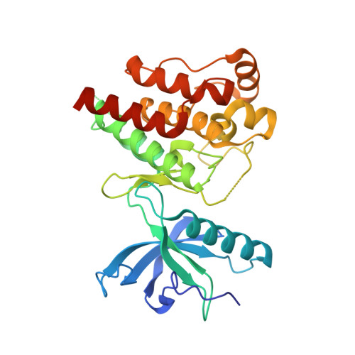
    6V9C

  • PubMed Abstract: 

    Hepatocellular carcinoma (HCC) accounts for a majority of primary liver cancer and is one of the most common forms of cancer worldwide. Aberrant signaling of the FGF19-FGFR4 pathway leads to HCC in mice and is hypothesized to be a driver in FGF19 amplified HCC in humans. Multiple small molecule inhibitors have been pursued as targeted therapies for HCC in recent years, including several selective FGFR4 inhibitors that are currently being evaluated in clinical trials. Herein, we report a novel series of highly selective, covalent 2-amino-6,8-dimethyl-pyrido[2,3- d ]pyrimidin-7(8 H )-ones that potently and selectively inhibit FGFR4 signaling through covalent modification of Cys552, which was confirmed by X-ray crystallography. Correlative target occupancy and pFGFR4 inhibition were observed in vivo, as well as tumor regression in preclinical models of orthotopic and sorafenib-resistant HCC.


  • Organizational Affiliation
    • Departments of Chemistry, Biology, and Biochemistry, Bristol Myers Squibb, 200 Cambridge Park Drive, Cambridge, Massachusetts 02140, United States.

Macromolecule Content 

  • Total Structure Weight: 34.58 kDa 
  • Atom Count: 2,421 
  • Modeled Residue Count: 279 
  • Deposited Residue Count: 300 
  • Unique protein chains: 1

Macromolecules

Find similar proteins by:|  3D Structure
Entity ID: 1
MoleculeChains  Sequence LengthOrganismDetailsImage
Fibroblast growth factor receptor 4300Homo sapiensMutation(s): 0 
Gene Names: FGFR4JTK2TKF
EC: 2.7.10.1
UniProt & NIH Common Fund Data Resources
Find proteins for P22455 (Homo sapiens)
Explore P22455 
Go to UniProtKB:  P22455
PHAROS:  P22455
GTEx:  ENSG00000160867 
Entity Groups
Sequence Clusters30% Identity50% Identity70% Identity90% Identity95% Identity100% Identity
UniProt GroupP22455
Sequence Annotations
Expand
Reference Sequence

Small Molecules

Ligands 2 Unique
IDChains Name / Formula / InChI Key2D Diagram3D Interactions
QS7
(Subject of Investigation/LOI)

Query on QS7


Download:Ideal Coordinates CCD File
B [auth A]N-[(3R,4S)-4-{[6-(2,6-dichloro-3,5-dimethoxyphenyl)-8-methyl-7-oxo-7,8-dihydropyrido[2,3-d]pyrimidin-2-yl]amino}oxolan-3-yl]prop-2-enamide
C23 H23 Cl2 N5 O5
ZIJVLVUPDVUSMA-UONOGXRCSA-N
SO4

Query on SO4


Download:Ideal Coordinates CCD File
C [auth A],
D [auth A]
SULFATE ION
O4 S
QAOWNCQODCNURD-UHFFFAOYSA-L
Binding Affinity Annotations 
IDSourceBinding Affinity
QS7 BindingDB:  6V9C IC50: min: 6.5, max: 500 (nM) from 4 assay(s)

Experimental Data & Validation

Experimental Data

  • Method: X-RAY DIFFRACTION
  • Resolution: 1.90 Å
  • R-Value Free:  0.227 (Depositor), 0.230 (DCC) 
  • R-Value Work:  0.199 (Depositor), 0.199 (DCC) 
  • R-Value Observed: 0.201 (Depositor) 
Space Group: P 1 21 1
Unit Cell:
Length ( Å )Angle ( ˚ )
a = 44.067α = 90
b = 58.67β = 97.57
c = 60.747γ = 90
Software Package:
Software NamePurpose
BUSTERrefinement
Aimlessdata scaling
PDB_EXTRACTdata extraction
XDSdata reduction
MOLREPphasing

Structure Validation

View Full Validation Report


Ligand Structure Quality Assessment 


Entry History 

Deposition Data

Revision History  (Full details and data files)

  • Version 1.0: 2020-03-25
    Type: Initial release
  • Version 1.1: 2020-11-04
    Changes: Database references
  • Version 1.2: 2023-10-11
    Changes: Data collection, Database references, Refinement description
  • Version 1.3: 2024-11-20
    Changes: Structure summary