6VBA | pdb_00006vba

Structure of human Uracil DNA Glycosylase (UDG) bound to Aurintricarboxylic acid (ATA)


Experimental Data Snapshot

  • Method: X-RAY DIFFRACTION
  • Resolution: 1.80 Å
  • R-Value Free: 
    0.234 (Depositor), 0.220 (DCC) 
  • R-Value Work: 
    0.179 (Depositor) 
  • R-Value Observed: 
    0.182 (Depositor) 

Starting Model: experimental
View more details

wwPDB Validation 3D Report Full Report

Validation slider image for 6VBA

Ligand Structure Quality Assessment 


This is version 1.3 of the entry. See complete history

Literature

An effective human uracil-DNA glycosylase inhibitor targets the open pre-catalytic active site conformation.

Nguyen, M.T.Moiani, D.Ahmed, Z.Arvai, A.S.Namjoshi, S.Shin, D.S.Fedorov, Y.Selvik, E.J.Jones, D.E.Pink, J.Yan, Y.Laverty, D.J.Nagel, Z.D.Tainer, J.A.Gerson, S.L.

(2021) Prog Biophys Mol Biol 163: 143-159

  • DOI: https://doi.org/10.1016/j.pbiomolbio.2021.02.004
  • Primary Citation Related Structures: 
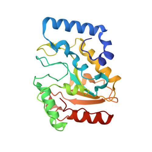
    6VBA

  • PubMed Abstract: 

    Human uracil DNA-glycosylase (UDG) is the prototypic and first identified DNA glycosylase with a vital role in removing deaminated cytosine and incorporated uracil and 5-fluorouracil (5-FU) from DNA. UDG depletion sensitizes cells to high APOBEC3B deaminase and to pemetrexed (PEM) and floxuridine (5-FdU), which are toxic to tumor cells through incorporation of uracil and 5-FU into DNA. To identify small-molecule UDG inhibitors for pre-clinical evaluation, we optimized biochemical screening of a selected diversity collection of >3,000 small-molecules. We found aurintricarboxylic acid (ATA) as an inhibitor of purified UDG at an initial calculated IC 50  < 100 nM. Subsequent enzymatic assays confirmed effective ATA inhibition but with an IC 50 of 700 nM and showed direct binding to the human UDG with a K D of <700 nM. ATA displays preferential, dose-dependent binding to purified human UDG compared to human 8-oxoguanine DNA glycosylase. ATA did not bind uracil-containing DNA at these concentrations. Yet, combined crystal structure and in silico docking results unveil ATA interactions with the DNA binding channel and uracil-binding pocket in an open, destabilized UDG conformation. Biologically relevant ATA inhibition of UDG was measured in cell lysates from human DLD1 colon cancer cells and in MCF-7 breast cancer cells using a host cell reactivation assay. Collective findings provide proof-of-principle for development of an ATA-based chemotype and "door stopper" strategy targeting inhibitor binding to a destabilized, open pre-catalytic glycosylase conformation that prevents active site closing for functional DNA binding and nucleotide flipping needed to excise altered bases in DNA.


  • Organizational Affiliation
    • Case Western Reserve University, Department of Biochemistry, Cleveland, OH, 44106, USA.

Macromolecule Content 

  • Total Structure Weight: 25.97 kDa 
  • Atom Count: 2,129 
  • Modeled Residue Count: 223 
  • Deposited Residue Count: 223 
  • Unique protein chains: 1

Macromolecules

Find similar proteins by:|  3D Structure
Entity ID: 1
MoleculeChains  Sequence LengthOrganismDetailsImage
Uracil-DNA glycosylase223Homo sapiensMutation(s): 0 
Gene Names: UNGDGUUNG1UNG15
EC: 3.2.2.27
UniProt & NIH Common Fund Data Resources
Find proteins for P13051 (Homo sapiens)
Explore P13051 
Go to UniProtKB:  P13051
PHAROS:  P13051
GTEx:  ENSG00000076248 
Entity Groups
Sequence Clusters30% Identity50% Identity70% Identity90% Identity95% Identity100% Identity
UniProt GroupP13051
Sequence Annotations
Expand
Reference Sequence

Small Molecules

Ligands 1 Unique
IDChains Name / Formula / InChI Key2D Diagram3D Interactions
QU4
(Subject of Investigation/LOI)

Query on QU4


Download:Ideal Coordinates CCD File
B [auth A]3,3'-[(3-carboxy-4-oxocyclohexa-2,5-dien-1-ylidene)methylene]bis(6-hydroxybenzoic acid)
C22 H14 O9
GIXWDMTZECRIJT-UHFFFAOYSA-N

Experimental Data & Validation

Experimental Data

  • Method: X-RAY DIFFRACTION
  • Resolution: 1.80 Å
  • R-Value Free:  0.234 (Depositor), 0.220 (DCC) 
  • R-Value Work:  0.179 (Depositor) 
  • R-Value Observed: 0.182 (Depositor) 
Space Group: P 21 21 2
Unit Cell:
Length ( Å )Angle ( ˚ )
a = 73.648α = 90
b = 54.775β = 90
c = 59.991γ = 90
Software Package:
Software NamePurpose
PHENIXrefinement
XDSdata reduction
XDSdata scaling
PHASERphasing

Structure Validation

View Full Validation Report


Ligand Structure Quality Assessment 


Entry History 

& Funding Information

Deposition Data


Funding OrganizationLocationGrant Number
National Institutes of Health/National Cancer Institute (NIH/NCI)United StatesR35CA220430
Robert A. Welch FoundationUnited StatesCFS127800-80-101399-50

Revision History  (Full details and data files)

  • Version 1.0: 2021-03-03
    Type: Initial release
  • Version 1.1: 2021-04-28
    Changes: Database references
  • Version 1.2: 2021-06-16
    Changes: Database references
  • Version 1.3: 2023-10-11
    Changes: Data collection, Database references, Refinement description