6VUM

Structure of nevanimibe-bound human tetrameric ACAT1


Experimental Data Snapshot

  • Method: ELECTRON MICROSCOPY
  • Resolution: 3.67 Å
  • Aggregation State: CELL 
  • Reconstruction Method: SINGLE PARTICLE 

wwPDB Validation   3D Report Full Report


This is version 1.2 of the entry. See complete history


Literature

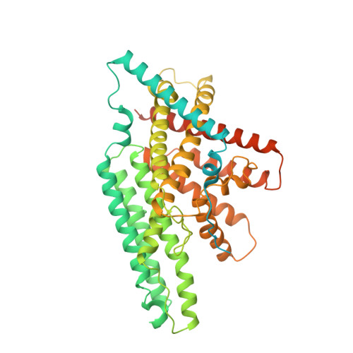
Structure of nevanimibe-bound tetrameric human ACAT1.

Long, T.Sun, Y.Hassan, A.Qi, X.Li, X.

(2020) Nature 581: 339-343

  • DOI: https://doi.org/10.1038/s41586-020-2295-8
  • Primary Citation of Related Structures:  
    6VUM

  • PubMed Abstract: 

    Cholesterol is an essential component of mammalian cell membranes, constituting up to 50% of plasma membrane lipids. By contrast, it accounts for only 5% of lipids in the endoplasmic reticulum (ER) 1 . The ER enzyme sterol O-acyltransferase 1 (also named acyl-coenzyme A:cholesterol acyltransferase, ACAT1) transfers a long-chain fatty acid to cholesterol to form cholesteryl esters that coalesce into cytosolic lipid droplets. Under conditions of cholesterol overload, ACAT1 maintains the low cholesterol concentration of the ER and thereby has an essential role in cholesterol homeostasis 2,3 . ACAT1 has also been implicated in Alzheimer's disease 4 , atherosclerosis 5 and cancers 6 . Here we report a cryo-electron microscopy structure of human ACAT1 in complex with nevanimibe 7 , an inhibitor that is in clinical trials for the treatment of congenital adrenal hyperplasia. The ACAT1 holoenzyme is a tetramer that consists of two homodimers. Each monomer contains nine transmembrane helices (TMs), six of which (TM4-TM9) form a cavity that accommodates nevanimibe and an endogenous acyl-coenzyme A. This cavity also contains a histidine that has previously been identified as essential for catalytic activity 8 . Our structural data and biochemical analyses provide a physical model to explain the process of cholesterol esterification, as well as details of the interaction between nevanimibe and ACAT1, which may help to accelerate the development of ACAT1 inhibitors to treat related diseases.


  • Organizational Affiliation

    Department of Molecular Genetics, University of Texas Southwestern Medical Center, Dallas, TX, USA.


Macromolecules
Find similar proteins by:  (by identity cutoff)  |  3D Structure
Entity ID: 1
MoleculeChains Sequence LengthOrganismDetailsImage
Sterol O-acyltransferase 1
A, B, C, D
558Homo sapiensMutation(s): 0 
Gene Names: SOAT1ACACTACACT1ACATACAT1SOATSTAT
EC: 2.3.1.26
Membrane Entity: Yes 
UniProt & NIH Common Fund Data Resources
Find proteins for P35610 (Homo sapiens)
Explore P35610 
Go to UniProtKB:  P35610
PHAROS:  P35610
GTEx:  ENSG00000057252 
Entity Groups  
Sequence Clusters30% Identity50% Identity70% Identity90% Identity95% Identity100% Identity
UniProt GroupP35610
Sequence Annotations
Expand
  • Reference Sequence
Small Molecules
Ligands 5 Unique
IDChains Name / Formula / InChI Key2D Diagram3D Interactions
3VV (Subject of Investigation/LOI)
Query on 3VV

Download Ideal Coordinates CCD File 
L [auth B],
W [auth D]
S-{(3R,5R,9R)-1-[(2R,3S,4R,5R)-5-(6-amino-9H-purin-9-yl)-4-hydroxy-3-(phosphonooxy)tetrahydrofuran-2-yl]-3,5,9-trihydroxy-8,8-dimethyl-3,5-dioxido-10,14-dioxo-2,4,6-trioxa-11,15-diaza-3lambda~5~,5lambda~5~-diphosphaheptadecan-17-yl} (9Z)-octadec-9-enethioate (non-preferred name)
C39 H68 N7 O17 P3 S
XDUHQPOXLUAVEE-BPMMELMSSA-N
COA
Query on COA

Download Ideal Coordinates CCD File 
J [auth A],
U [auth C]
COENZYME A
C21 H36 N7 O16 P3 S
RGJOEKWQDUBAIZ-IBOSZNHHSA-N
ROV (Subject of Investigation/LOI)
Query on ROV

Download Ideal Coordinates CCD File 
I [auth A],
O [auth B],
T [auth C],
Z [auth D]
nevanimibe
C27 H39 N3 O
PKKNCEXEVUFFFI-UHFFFAOYSA-N
CLR
Query on CLR

Download Ideal Coordinates CCD File 
F [auth A]
G [auth A]
H [auth A]
M [auth B]
N [auth B]
F [auth A],
G [auth A],
H [auth A],
M [auth B],
N [auth B],
Q [auth C],
R [auth C],
S [auth C],
X [auth D],
Y [auth D]
CHOLESTEROL
C27 H46 O
HVYWMOMLDIMFJA-DPAQBDIFSA-N
OLA
Query on OLA

Download Ideal Coordinates CCD File 
E [auth A],
K [auth B],
P [auth C],
V [auth D]
OLEIC ACID
C18 H34 O2
ZQPPMHVWECSIRJ-KTKRTIGZSA-N
Experimental Data & Validation

Experimental Data

  • Method: ELECTRON MICROSCOPY
  • Resolution: 3.67 Å
  • Aggregation State: CELL 
  • Reconstruction Method: SINGLE PARTICLE 

Structure Validation

View Full Validation Report


Entry History 

Deposition Data

Revision History  (Full details and data files)

  • Version 1.0: 2020-05-13
    Type: Initial release
  • Version 1.1: 2020-06-03
    Changes: Database references
  • Version 1.2: 2024-03-06
    Changes: Data collection, Database references