6W0M | pdb_00006w0m

Human 8-oxoguanine glycosylase crosslinked with oxoG lesion containing DNA


Experimental Data Snapshot

  • Method: X-RAY DIFFRACTION
  • Resolution: 2.37 Å
  • R-Value Free: 
    0.266 (Depositor), 0.266 (DCC) 
  • R-Value Work: 
    0.197 (Depositor), 0.203 (DCC) 
  • R-Value Observed: 
    0.200 (Depositor) 

Starting Model: experimental
View more details

wwPDB Validation 3D Report Full Report

Validation slider image for 6W0M

Ligand Structure Quality Assessment 


This is version 1.1 of the entry. See complete history

Literature

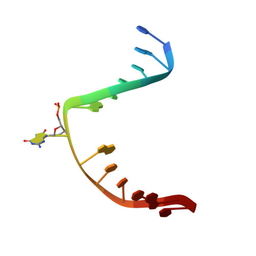
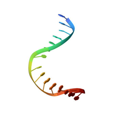
The trajectory of intrahelical lesion recognition and extrusion by the human 8-oxoguanine DNA glycosylase.

Shigdel, U.K.Ovchinnikov, V.Lee, S.J.Shih, J.A.Karplus, M.Nam, K.Verdine, G.L.

(2020) Nat Commun 11: 4437-4437

  • DOI: https://doi.org/10.1038/s41467-020-18290-2
  • Primary Citation Related Structures: 
    6W0M, 6W0R, 6W13

  • PubMed Abstract: 

    Efficient search for DNA damage embedded in vast expanses of the DNA genome presents one of the greatest challenges to DNA repair enzymes. We report here crystal structures of human 8-oxoguanine (oxoG) DNA glycosylase, hOGG1, that interact with the DNA containing the damaged base oxoG and the normal base G while they are nested in the DNA helical stack. The structures reveal that hOGG1 engages the DNA using different protein-DNA contacts from those observed in the previously determined lesion recognition complex and other hOGG1-DNA complexes. By applying molecular dynamics simulations, we have determined the pathways taken by the lesion and normal bases when extruded from the DNA helix and their associated free energy profiles. These results reveal how the human oxoG DNA glycosylase hOGG1 locates the lesions inside the DNA helix and facilitates their extrusion for repair.


  • Organizational Affiliation
    • Department of Stem Cell and Regenerative Biology, Harvard University, Cambridge, MA, 02138, USA.

Macromolecule Content 

  • Total Structure Weight: 44.36 kDa 
  • Atom Count: 3,181 
  • Modeled Residue Count: 342 
  • Deposited Residue Count: 345 
  • Unique protein chains: 1
  • Unique nucleic acid chains: 2

Macromolecules


Find similar proteins by:|  3D Structure
Entity ID: 1
MoleculeChains  Sequence LengthOrganismDetailsImage
N-glycosylase/DNA lyase317Homo sapiensMutation(s): 3 
Gene Names: OGG1MMHMUTMOGH1
EC: 3.2.2 (PDB Primary Data), 4.2.99.18 (PDB Primary Data)
UniProt & NIH Common Fund Data Resources
Find proteins for O15527 (Homo sapiens)
Explore O15527 
Go to UniProtKB:  O15527
PHAROS:  O15527
GTEx:  ENSG00000114026 
Entity Groups
Sequence Clusters30% Identity50% Identity70% Identity90% Identity95% Identity100% Identity
UniProt GroupO15527
Sequence Annotations
Expand
Reference Sequence
Find similar nucleic acids by:  Sequence
Entity ID: 2
MoleculeChains LengthOrganismImage
DNA (5'-D(P*CP*GP*TP*CP*CP*AP*(8OG)P*GP*TP*CP*TP*AP*C)-3')13Homo sapiens
Sequence Annotations
Expand
Reference Sequence
Find similar nucleic acids by:  Sequence
Entity ID: 3
MoleculeChains LengthOrganismImage
DNA (5'-D(P*GP*GP*TP*AP*GP*AP*CP*CP*TP*GP*GP*AP*CP*GP*C)-3')15Homo sapiens
Sequence Annotations
Expand
Reference Sequence

Experimental Data & Validation

Experimental Data

  • Method: X-RAY DIFFRACTION
  • Resolution: 2.37 Å
  • R-Value Free:  0.266 (Depositor), 0.266 (DCC) 
  • R-Value Work:  0.197 (Depositor), 0.203 (DCC) 
  • R-Value Observed: 0.200 (Depositor) 
Space Group: P 65 2 2
Unit Cell:
Length ( Å )Angle ( ˚ )
a = 91.678α = 90
b = 91.678β = 90
c = 212.106γ = 120
Software Package:
Software NamePurpose
REFMACrefinement
HKL-2000data scaling
PDB_EXTRACTdata extraction
PHASERphasing
HKL-2000data collection
HKL-2000data processing

Structure Validation

View Full Validation Report


Ligand Structure Quality Assessment 


Entry History 

& Funding Information

Deposition Data


Funding OrganizationLocationGrant Number
National Institutes of Health/National Cancer Institute (NIH/NCI)United States--

Revision History  (Full details and data files)

  • Version 1.0: 2020-09-23
    Type: Initial release
  • Version 1.1: 2023-10-11
    Changes: Advisory, Data collection, Database references, Refinement description