6WB8

Cryo-EM structure of PKD2 C331S disease variant


Experimental Data Snapshot

  • Method: ELECTRON MICROSCOPY
  • Resolution: 3.24 Å
  • Aggregation State: PARTICLE 
  • Reconstruction Method: SINGLE PARTICLE 

wwPDB Validation   3D Report Full Report


This is version 1.4 of the entry. See complete history


Literature

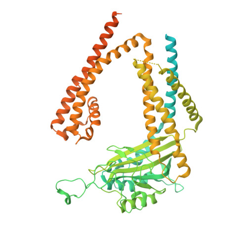
Molecular dysregulation of ciliary polycystin-2 channels caused by variants in the TOP domain.

Vien, T.N.Wang, J.Ng, L.C.T.Cao, E.DeCaen, P.G.

(2020) Proc Natl Acad Sci U S A 117: 10329-10338

  • DOI: https://doi.org/10.1073/pnas.1920777117
  • Primary Citation of Related Structures:  
    6WB8

  • PubMed Abstract: 

    Genetic variants in PKD2 which encodes for the polycystin-2 ion channel are responsible for many clinical cases of autosomal dominant polycystic kidney disease (ADPKD). Despite our strong understanding of the genetic basis of ADPKD, we do not know how most variants impact channel function. Polycystin-2 is found in organelle membranes, including the primary cilium-an antennae-like structure on the luminal side of the collecting duct. In this study, we focus on the structural and mechanistic regulation of polycystin-2 by its TOP domain-a site with unknown function that is commonly altered by missense variants. We use direct cilia electrophysiology, cryogenic electron microscopy, and superresolution imaging to determine that variants of the TOP domain finger 1 motif destabilizes the channel structure and impairs channel opening without altering cilia localization and channel assembly. Our findings support the channelopathy classification of PKD2 variants associated with ADPKD, where polycystin-2 channel dysregulation in the primary cilia may contribute to cystogenesis.


  • Organizational Affiliation

    Department of Pharmacology, Feinberg School of Medicine, Northwestern University, Chicago, IL 60611.


Macromolecules
Find similar proteins by:  (by identity cutoff)  |  3D Structure
Entity ID: 1
MoleculeChains Sequence LengthOrganismDetailsImage
Polycystin-2
A, B, C, D
756Homo sapiensMutation(s): 1 
Gene Names: PKD2TRPP2
Membrane Entity: Yes 
UniProt & NIH Common Fund Data Resources
Find proteins for Q13563 (Homo sapiens)
Explore Q13563 
Go to UniProtKB:  Q13563
PHAROS:  Q13563
GTEx:  ENSG00000118762 
Entity Groups  
Sequence Clusters30% Identity50% Identity70% Identity90% Identity95% Identity100% Identity
UniProt GroupQ13563
Sequence Annotations
Expand
  • Reference Sequence
Small Molecules
Ligands 1 Unique
IDChains Name / Formula / InChI Key2D Diagram3D Interactions
NAG (Subject of Investigation/LOI)
Query on NAG

Download Ideal Coordinates CCD File 
E [auth A]
F [auth A]
G [auth A]
H [auth B]
I [auth B]
E [auth A],
F [auth A],
G [auth A],
H [auth B],
I [auth B],
J [auth B],
K [auth C],
L [auth C],
M [auth C],
N [auth D],
O [auth D],
P [auth D]
2-acetamido-2-deoxy-beta-D-glucopyranose
C8 H15 N O6
OVRNDRQMDRJTHS-FMDGEEDCSA-N
Experimental Data & Validation

Experimental Data

  • Method: ELECTRON MICROSCOPY
  • Resolution: 3.24 Å
  • Aggregation State: PARTICLE 
  • Reconstruction Method: SINGLE PARTICLE 

Structure Validation

View Full Validation Report


Entry History & Funding Information

Deposition Data


Funding OrganizationLocationGrant Number
National Institutes of Health/National Institute of Diabetes and Digestive and Kidney Disease (NIH/NIDDK)United StatesR01 DK110575

Revision History  (Full details and data files)

  • Version 1.0: 2020-04-22
    Type: Initial release
  • Version 1.1: 2020-05-06
    Changes: Database references
  • Version 1.2: 2020-05-13
    Changes: Database references
  • Version 1.3: 2020-05-27
    Changes: Database references
  • Version 1.4: 2020-07-29
    Type: Remediation
    Reason: Carbohydrate remediation
    Changes: Data collection, Derived calculations, Structure summary