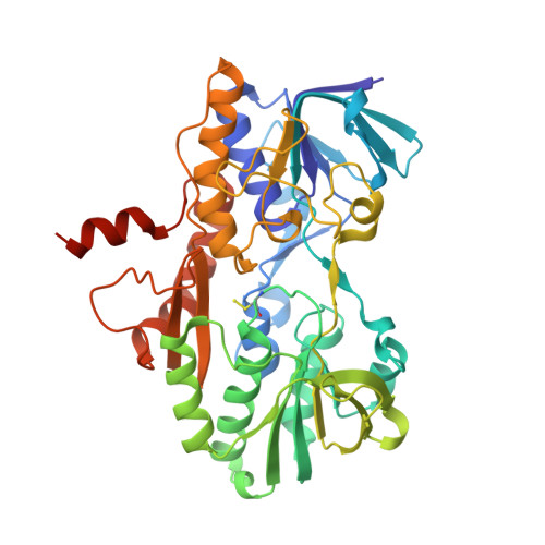
6WH6

Crystal structure of human sulfide quinone oxidoreductase in complex with coenzyme Q (cyanide soaked)


Experimental Data Snapshot

  • Method: X-RAY DIFFRACTION
  • Resolution: 2.25 Å
  • R-Value Free: 0.216 
  • R-Value Work: 0.172 
  • R-Value Observed: 0.174 

Starting Model: experimental
View more details

wwPDB Validation   3D Report Full Report


Ligand Structure Quality Assessment 


This is version 1.4 of the entry. See complete history


Literature

Dismantling and Rebuilding the Trisulfide Cofactor Demonstrates Its Essential Role in Human Sulfide Quinone Oxidoreductase.

Landry, A.P.Moon, S.Bonanata, J.Cho, U.S.Coitino, E.L.Banerjee, R.

(2020) J Am Chem Soc 142: 14295-14306

  • DOI: https://doi.org/10.1021/jacs.0c06066
  • Primary Citation of Related Structures:  
    6WH6

  • PubMed Abstract: 

    Sulfide quinone oxidoreductase (SQOR) catalyzes the first step in sulfide clearance, coupling H 2 S oxidation to coenzyme Q reduction. Recent structures of human SQOR revealed a sulfur atom bridging the SQOR active site cysteines in a trisulfide configuration. Here, we assessed the importance of this cofactor using kinetic, crystallographic, and computational modeling approaches. Cyanolysis of SQOR proceeds via formation of an intense charge transfer complex that subsequently decays to eliminate thiocyanate. We captured a disulfanyl-methanimido thioate intermediate in the SQOR crystal structure, revealing how cyanolysis leads to reversible loss of SQOR activity that is restored in the presence of sulfide. Computational modeling and MD simulations revealed an ∼10 5 -fold rate enhancement for nucleophilic addition of sulfide into the trisulfide versus a disulfide cofactor. The cysteine trisulfide in SQOR is thus critical for activity and provides a significant catalytic advantage over a cysteine disulfide.


  • Organizational Affiliation

    Department of Biological Chemistry, University of Michigan Medical School, Ann Arbor, Michigan 48109, United States.


Macromolecules
Find similar proteins by:  (by identity cutoff)  |  3D Structure
Entity ID: 1
MoleculeChains Sequence LengthOrganismDetailsImage
Sulfide:quinone oxidoreductase, mitochondrial
A, B
418Homo sapiensMutation(s): 0 
Gene Names: SQORSQRDLCGI-44
EC: 1.8.5 (PDB Primary Data), 1.8.5.8 (UniProt)
UniProt & NIH Common Fund Data Resources
Find proteins for Q9Y6N5 (Homo sapiens)
Explore Q9Y6N5 
Go to UniProtKB:  Q9Y6N5
PHAROS:  Q9Y6N5
GTEx:  ENSG00000137767 
Entity Groups  
Sequence Clusters30% Identity50% Identity70% Identity90% Identity95% Identity100% Identity
UniProt GroupQ9Y6N5
Sequence Annotations
Expand
  • Reference Sequence
Small Molecules
Modified Residues  1 Unique
IDChains TypeFormula2D DiagramParent
CSS
Query on CSS
A, B
L-PEPTIDE LINKINGC3 H7 N O2 S2CYS
Experimental Data & Validation

Experimental Data

  • Method: X-RAY DIFFRACTION
  • Resolution: 2.25 Å
  • R-Value Free: 0.216 
  • R-Value Work: 0.172 
  • R-Value Observed: 0.174 
  • Space Group: P 21 21 21
Unit Cell:
Length ( Å )Angle ( ˚ )
a = 78.393α = 90
b = 111.746β = 90
c = 134.046γ = 90
Software Package:
Software NamePurpose
REFMACrefinement
PHENIXrefinement
HKL-2000data reduction
HKL-2000data scaling
PHENIXphasing

Structure Validation

View Full Validation Report


Ligand Structure Quality Assessment 


Entry History & Funding Information

Deposition Data


Funding OrganizationLocationGrant Number
National Institutes of Health/National Institute of General Medical Sciences (NIH/NIGMS)United StatesGM130183

Revision History  (Full details and data files)

  • Version 1.0: 2021-04-21
    Type: Initial release
  • Version 1.1: 2021-11-03
    Changes: Database references, Derived calculations
  • Version 1.2: 2023-10-18
    Changes: Data collection, Refinement description
  • Version 1.3: 2023-11-15
    Changes: Data collection
  • Version 1.4: 2024-10-09
    Changes: Structure summary