6WKW

EM structure of CtBP2 with a minimal dehydrogenase domain of CtBP2

  • Classification: GENE REGULATION
  • Organism(s): Homo sapiens
  • Expression System: Escherichia coli
  • Mutation(s): No 

  • Deposited: 2020-04-17 Released: 2020-12-02 
  • Deposition Author(s): Jecrois, A.M.
  • Funding Organization(s): National Institutes of Health/National Institute of General Medical Sciences (NIH/NIGMS)

Experimental Data Snapshot

  • Method: ELECTRON MICROSCOPY
  • Resolution: 3.60 Å
  • Aggregation State: PARTICLE 
  • Reconstruction Method: SINGLE PARTICLE 

wwPDB Validation   3D Report Full Report


This is version 1.3 of the entry. See complete history


Literature

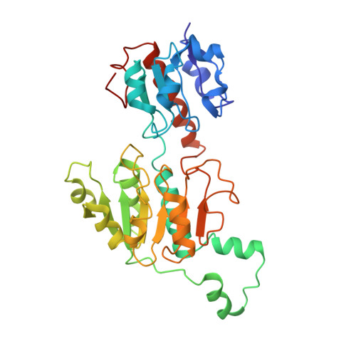
Cryo-EM structure of CtBP2 confirms tetrameric architecture.

Jecrois, A.M.Dcona, M.M.Deng, X.Bandyopadhyay, D.Grossman, S.R.Schiffer, C.A.Royer Jr., W.E.

(2021) Structure 29: 310

  • DOI: https://doi.org/10.1016/j.str.2020.11.008
  • Primary Citation of Related Structures:  
    6WKW

  • PubMed Abstract: 

    C-terminal binding proteins 1 and 2 (CtBP1 and CtBP2) are transcriptional regulators that activate or repress many genes involved in cellular development, apoptosis, and metastasis. NADH-dependent CtBP activation has been implicated in multiple types of cancer and poor patient prognosis. Central to understanding activation of CtBP in oncogenesis is uncovering how NADH triggers protein assembly, what level of assembly occurs, and if oncogenic activity depends upon such assembly. Here, we present the cryoelectron microscopic structures of two different constructs of CtBP2 corroborating that the native state of CtBP2 in the presence of NADH is tetrameric. The physiological relevance of the observed tetramer was demonstrated in cell culture, showing that CtBP tetramer-destabilizing mutants are defective for cell migration, transcriptional repression of E-cadherin, and activation of TIAM1. Together with our cryoelectron microscopy studies, these results highlight the tetramer as the functional oligomeric form of CtBP2.


  • Organizational Affiliation

    Department of Biochemistry and Molecular Pharmacology, University of Massachusetts Medical School, Worcester, MA 01605, USA.


Macromolecules
Find similar proteins by:  (by identity cutoff)  |  3D Structure
Entity ID: 1
MoleculeChains Sequence LengthOrganismDetailsImage
C-terminal-binding protein 2A [auth D],
B [auth C],
C [auth B],
D [auth A]
330Homo sapiensMutation(s): 0 
Gene Names: CTBP2
UniProt & NIH Common Fund Data Resources
Find proteins for P56545 (Homo sapiens)
Explore P56545 
Go to UniProtKB:  P56545
PHAROS:  P56545
GTEx:  ENSG00000175029 
Entity Groups  
Sequence Clusters30% Identity50% Identity70% Identity90% Identity95% Identity100% Identity
UniProt GroupP56545
Sequence Annotations
Expand
  • Reference Sequence
Experimental Data & Validation

Experimental Data

  • Method: ELECTRON MICROSCOPY
  • Resolution: 3.60 Å
  • Aggregation State: PARTICLE 
  • Reconstruction Method: SINGLE PARTICLE 
EM Software:
TaskSoftware PackageVersion
RECONSTRUCTIONRELION3.0.2
MODEL REFINEMENTCoot

Structure Validation

View Full Validation Report


Entry History & Funding Information

Deposition Data


Funding OrganizationLocationGrant Number
National Institutes of Health/National Institute of General Medical Sciences (NIH/NIGMS)United StatesR01 GM119014

Revision History  (Full details and data files)

  • Version 1.0: 2020-12-02
    Type: Initial release
  • Version 1.1: 2020-12-16
    Changes: Database references
  • Version 1.2: 2021-04-14
    Changes: Database references
  • Version 1.3: 2024-03-06
    Changes: Data collection, Database references, Refinement description