6WYD | pdb_00006wyd

CRYSTAL STRUCTURE OF MYELOPEROXIDASE SUBFORM C (MPO) COMPLEX WITH Compound-12 (AKA; 7-benzyl-1H-[1,2,3]triazolo[4,5-b]pyrid


Experimental Data Snapshot

  • Method: X-RAY DIFFRACTION
  • Resolution: 2.55 Å
  • R-Value Free: 
    0.254 (Depositor), 0.210 (DCC) 
  • R-Value Work: 
    0.194 (Depositor), 0.207 (DCC) 
  • R-Value Observed: 
    0.197 (Depositor) 

Starting Model: experimental
View more details

wwPDB Validation 3D Report Full Report

Validation slider image for 6WYD

Ligand Structure Quality Assessment 


This is version 1.2 of the entry. See complete history

Literature

Discovery and structure activity relationships of 7-benzyl triazolopyridines as stable, selective, and reversible inhibitors of myeloperoxidase.

Shaw, S.A.Vokits, B.P.Dilger, A.K.Viet, A.Clark, C.G.Abell, L.M.Locke, G.A.Duke, G.Kopcho, L.M.Dongre, A.Gao, J.Krishnakumar, A.Jusuf, S.Khan, J.Spronk, S.A.Basso, M.D.Zhao, L.Cantor, G.H.Onorato, J.M.Wexler, R.R.Duclos, F.Kick, E.K.

(2020) Bioorg Med Chem 28: 115723-115723

  • DOI: https://doi.org/10.1016/j.bmc.2020.115723
  • Primary Citation Related Structures: 
    6WXZ, 6WY0, 6WY5, 6WY7, 6WYD

  • PubMed Abstract: 

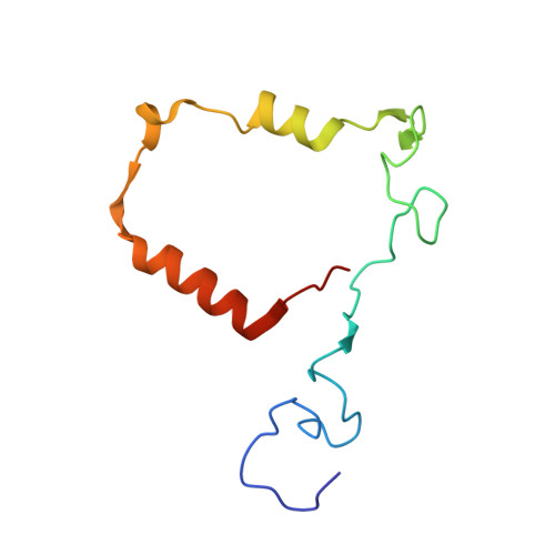
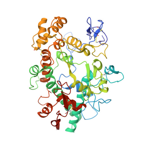
    Myeloperoxidase (MPO) is a heme peroxidase found in neutrophils, monocytes and macrophages that efficiently catalyzes the oxidation of endogenous chloride into hypochlorous acid for antimicrobial activity. Chronic MPO activation can lead to indiscriminate protein modification causing tissue damage, and has been associated with chronic inflammatory diseases, atherosclerosis, and acute cardiovascular events. Triazolopyrimidine 5 is a reversible MPO inhibitor; however it suffers from poor stability in acid, and is an irreversible inhibitor of the DNA repair protein methyl guanine methyl transferase (MGMT). Structure-based drug design was employed to discover benzyl triazolopyridines with improved MPO potency, as well as acid stability, no reactivity with MGMT, and selectivity against thyroid peroxidase (TPO). Structure-activity relationships, a crystal structure of the MPO-inhibitor complex, and acute in vivo pharmacodynamic data are described herein.


  • Organizational Affiliation
    • Bristol Myers Squibb Company, P.O. Box 5400, Princeton, NJ 08543-5400, United States. Electronic address: scott.shaw@bms.com.

Macromolecule Content 

  • Total Structure Weight: 271.96 kDa 
  • Atom Count: 18,734 
  • Modeled Residue Count: 2,272 
  • Deposited Residue Count: 2,288 
  • Unique protein chains: 2

Macromolecules

Find similar proteins by:|  3D Structure
Entity ID: 1
MoleculeChains  Sequence LengthOrganismDetailsImage
Myeloperoxidase light chainA,
C [auth D],
E [auth F],
G [auth H]
105Homo sapiensMutation(s): 0 
EC: 1.11.2.2
UniProt & NIH Common Fund Data Resources
Find proteins for P05164 (Homo sapiens)
Explore P05164 
Go to UniProtKB:  P05164
PHAROS:  P05164
GTEx:  ENSG00000005381 
Entity Groups
Sequence Clusters30% Identity50% Identity70% Identity90% Identity95% Identity100% Identity
UniProt GroupP05164
Sequence Annotations
Expand
Reference Sequence
Find similar proteins by:|  3D Structure
Entity ID: 2
MoleculeChains  Sequence LengthOrganismDetailsImage
Myeloperoxidase heavy chainB,
D [auth E],
F [auth G],
H [auth I]
467Homo sapiensMutation(s): 0 
EC: 1.11.2.2
UniProt & NIH Common Fund Data Resources
Find proteins for P05164 (Homo sapiens)
Explore P05164 
Go to UniProtKB:  P05164
PHAROS:  P05164
GTEx:  ENSG00000005381 
Entity Groups
Sequence Clusters30% Identity50% Identity70% Identity90% Identity95% Identity100% Identity
UniProt GroupP05164
Sequence Annotations
Expand
Reference Sequence

Small Molecules

Ligands 8 Unique
IDChains Name / Formula / InChI Key2D Diagram3D Interactions
HEC

Query on HEC


Download:Ideal Coordinates CCD File
FA [auth E],
IB [auth I],
O [auth B],
PA [auth G]
HEME C
C34 H34 Fe N4 O4
HXQIYSLZKNYNMH-LJNAALQVSA-N
7GD
(Subject of Investigation/LOI)

Query on 7GD


Download:Ideal Coordinates CCD File
GA [auth E],
T [auth B],
UA [auth G],
XA [auth H]
7-benzyl-1H-[1,2,3]triazolo[4,5-b]pyridin-5-amine
C12 H11 N5
SGFFXIGIHATQNC-UHFFFAOYSA-N
NAG

Query on NAG


Download:Ideal Coordinates CCD File
AA [auth E]
BA [auth E]
CA [auth E]
DA [auth E]
DB [auth I]
AA [auth E],
BA [auth E],
CA [auth E],
DA [auth E],
DB [auth I],
EB [auth I],
FB [auth I],
GB [auth I],
J [auth B],
K [auth B],
L [auth B],
LA [auth G],
M [auth B],
MA [auth G],
NA [auth G],
P [auth B],
QA [auth G],
W [auth E],
ZA [auth I]
2-acetamido-2-deoxy-beta-D-glucopyranose
C8 H15 N O6
OVRNDRQMDRJTHS-FMDGEEDCSA-N
BMA

Query on BMA


Download:Ideal Coordinates CCD File
AB [auth I],
Q [auth B],
RA [auth G],
X [auth E]
beta-D-mannopyranose
C6 H12 O6
WQZGKKKJIJFFOK-RWOPYEJCSA-N
MAN

Query on MAN


Download:Ideal Coordinates CCD File
BB [auth I]
CB [auth I]
R [auth B]
S [auth B]
SA [auth G]
BB [auth I],
CB [auth I],
R [auth B],
S [auth B],
SA [auth G],
TA [auth G],
Y [auth E],
Z [auth E]
alpha-D-mannopyranose
C6 H12 O6
WQZGKKKJIJFFOK-PQMKYFCFSA-N
FUC

Query on FUC


Download:Ideal Coordinates CCD File
EA [auth E],
HB [auth I],
N [auth B],
OA [auth G]
alpha-L-fucopyranose
C6 H12 O5
SHZGCJCMOBCMKK-SXUWKVJYSA-N
CA

Query on CA


Download:Ideal Coordinates CCD File
HA [auth E],
JB [auth I],
U [auth B],
VA [auth G]
CALCIUM ION
Ca
BHPQYMZQTOCNFJ-UHFFFAOYSA-N
CL

Query on CL


Download:Ideal Coordinates CCD File
I [auth A]
IA [auth E]
JA [auth E]
KA [auth F]
KB [auth I]
I [auth A],
IA [auth E],
JA [auth E],
KA [auth F],
KB [auth I],
V [auth B],
WA [auth G],
YA [auth H]
CHLORIDE ION
Cl
VEXZGXHMUGYJMC-UHFFFAOYSA-M

Experimental Data & Validation

Experimental Data

  • Method: X-RAY DIFFRACTION
  • Resolution: 2.55 Å
  • R-Value Free:  0.254 (Depositor), 0.210 (DCC) 
  • R-Value Work:  0.194 (Depositor), 0.207 (DCC) 
  • R-Value Observed: 0.197 (Depositor) 
Space Group: C 2 2 21
Unit Cell:
Length ( Å )Angle ( ˚ )
a = 143.683α = 90
b = 150.651β = 90
c = 231.06γ = 90
Software Package:
Software NamePurpose
BUSTERrefinement
PDB_EXTRACTdata extraction
XDSdata reduction
Aimlessdata scaling
PHASERphasing

Structure Validation

View Full Validation Report


Ligand Structure Quality Assessment 


Entry History 

Deposition Data

  • Released Date: 2020-10-14 
  • Deposition Author(s): Khan, J.A.

Revision History  (Full details and data files)

  • Version 1.0: 2020-10-14
    Type: Initial release
  • Version 1.1: 2023-10-18
    Changes: Advisory, Data collection, Database references, Refinement description
  • Version 1.2: 2024-10-23
    Changes: Structure summary