6XQY

Crystal structure of the catalytic domain of PBP2 S310A from Neisseria gonorrhoeae at pH 9.5


Experimental Data Snapshot

  • Method: X-RAY DIFFRACTION
  • Resolution: 1.90 Å
  • R-Value Free: 0.195 
  • R-Value Work: 0.165 
  • R-Value Observed: 0.166 

wwPDB Validation   3D Report Full Report


This is version 1.2 of the entry. See complete history


Literature

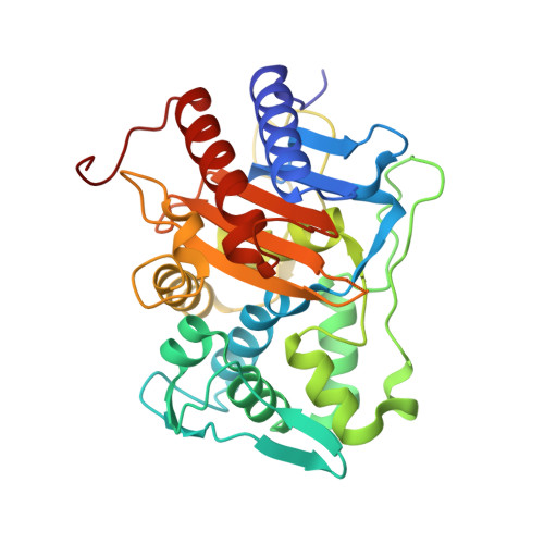
Mutations in PBP2 from ceftriaxone-resistant Neisseria gonorrhoeae alter the dynamics of the beta 3-beta 4 loop to favor a low-affinity drug-binding state.

Fenton, B.A.Tomberg, J.Sciandra, C.A.Nicholas, R.A.Davies, C.Zhou, P.

(2021) J Biol Chem 297: 101188-101188

  • DOI: https://doi.org/10.1016/j.jbc.2021.101188
  • Primary Citation of Related Structures:  
    6XQV, 6XQX, 6XQY, 6XQZ

  • PubMed Abstract: 

    Resistance to the extended-spectrum cephalosporin ceftriaxone in the pathogenic bacteria Neisseria gonorrhoeae is conferred by mutations in penicillin-binding protein 2 (PBP2), the lethal target of the antibiotic, but how these mutations exert their effect at the molecular level is unclear. Using solution NMR, X-ray crystallography, and isothermal titration calorimetry, we report that WT PBP2 exchanges dynamically between a low-affinity state with an extended β3-β4 loop conformation and a high-affinity state with an inward β3-β4 loop conformation. Histidine-514, which is located at the boundary of the β4 strand, plays an important role during the exchange between these two conformational states. We also find that mutations present in PBP2 from H041, a ceftriaxone-resistant strain of N. gonorrhoeae, increase resistance to ceftriaxone by destabilizing the inward β3-β4 loop conformation or stabilizing the extended β3-β4 loop conformation to favor the low-affinity drug-binding state. These observations reveal a unique mechanism for ceftriaxone resistance, whereby mutations in PBP2 lower the proportion of target molecules in the high-affinity drug-binding state and thus reduce inhibition at lower drug concentrations.


  • Organizational Affiliation

    Department of Biochemistry, Duke University School of Medicine, Durham, North Carolina, USA.


Macromolecules
Find similar proteins by:  (by identity cutoff)  |  3D Structure
Entity ID: 1
MoleculeChains Sequence LengthOrganismDetailsImage
Probable peptidoglycan D,D-transpeptidase PenA
A, B
327Neisseria gonorrhoeaeMutation(s): 1 
Gene Names: penA
EC: 3.4.16.4
UniProt
Find proteins for P08149 (Neisseria gonorrhoeae)
Explore P08149 
Go to UniProtKB:  P08149
Entity Groups  
Sequence Clusters30% Identity50% Identity70% Identity90% Identity95% Identity100% Identity
UniProt GroupP08149
Sequence Annotations
Expand
  • Reference Sequence
Small Molecules
Ligands 1 Unique
IDChains Name / Formula / InChI Key2D Diagram3D Interactions
CL
Query on CL

Download Ideal Coordinates CCD File 
C [auth B]CHLORIDE ION
Cl
VEXZGXHMUGYJMC-UHFFFAOYSA-M
Experimental Data & Validation

Experimental Data

  • Method: X-RAY DIFFRACTION
  • Resolution: 1.90 Å
  • R-Value Free: 0.195 
  • R-Value Work: 0.165 
  • R-Value Observed: 0.166 
  • Space Group: P 1 21 1
Unit Cell:
Length ( Å )Angle ( ˚ )
a = 44.94α = 90
b = 76.55β = 91.636
c = 87.9γ = 90
Software Package:
Software NamePurpose
XDSdata reduction
XSCALEdata scaling
PHASERphasing
PHENIXrefinement

Structure Validation

View Full Validation Report


Entry History & Funding Information

Deposition Data


Funding OrganizationLocationGrant Number
National Institutes of Health/National Institute of General Medical Sciences (NIH/NIGMS)United StatesGM066861

Revision History  (Full details and data files)

  • Version 1.0: 2021-07-21
    Type: Initial release
  • Version 1.1: 2022-02-02
    Changes: Database references
  • Version 1.2: 2023-10-18
    Changes: Data collection, Refinement description