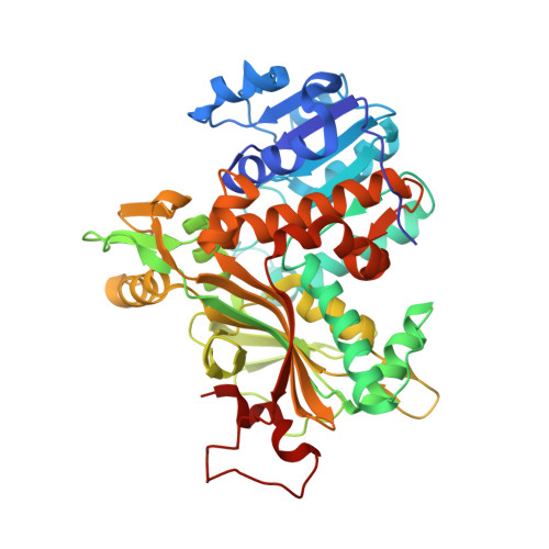
6Y87

Crystal Structure of the Homospermidine Synthase (HSS) from Pseudomonas aeruginosa in Complex with NAD and PUT


Experimental Data Snapshot

  • Method: X-RAY DIFFRACTION
  • Resolution: 2.15 Å
  • R-Value Free: 0.244 
  • R-Value Work: 0.203 
  • R-Value Observed: 0.205 

Starting Model: experimental
View more details

wwPDB Validation   3D Report Full Report


Ligand Structure Quality Assessment 


This is version 1.2 of the entry. See complete history


Literature

Structural and catalytic characterization of Blastochloris viridis and Pseudomonas aeruginosa homospermidine synthases supports the essential role of cation-pi interaction.

Helfrich, F.Scheidig, A.J.

(2021) Acta Crystallogr D Struct Biol 77: 1317-1335

  • DOI: https://doi.org/10.1107/S2059798321008937
  • Primary Citation of Related Structures:  
    6S3X, 6S49, 6S4D, 6S6G, 6S72, 6SEP, 6Y87

  • PubMed Abstract: 

    Polyamines influence medically relevant processes in the opportunistic pathogen Pseudomonas aeruginosa, including virulence, biofilm formation and susceptibility to antibiotics. Although homospermidine synthase (HSS) is part of the polyamine metabolism in various strains of P. aeruginosa, neither its role nor its structure has been examined so far. The reaction mechanism of the nicotinamide adenine dinucleotide (NAD + )-dependent bacterial HSS has previously been characterized based on crystal structures of Blastochloris viridis HSS (BvHSS). This study presents the crystal structure of P. aeruginosa HSS (PaHSS) in complex with its substrate putrescine. A high structural similarity between PaHSS and BvHSS with conservation of the catalytically relevant residues is demonstrated, qualifying BvHSS as a model for mechanistic studies of PaHSS. Following this strategy, crystal structures of single-residue variants of BvHSS are presented together with activity assays of PaHSS, BvHSS and BvHSS variants. For efficient homospermidine production, acidic residues are required at the entrance to the binding pocket (`ionic slide') and near the active site (`inner amino site') to attract and bind the substrate putrescine via salt bridges. The tryptophan residue at the active site stabilizes cationic reaction components by cation-π interaction, as inferred from the interaction geometry between putrescine and the indole ring plane. Exchange of this tryptophan for other amino acids suggests a distinct catalytic requirement for an aromatic interaction partner with a highly negative electrostatic potential. These findings substantiate the structural and mechanistic knowledge on bacterial HSS, a potential target for antibiotic design.


  • Organizational Affiliation

    Zoological Institute, University of Kiel, Am Botanischen Garten 1-9, 24118 Kiel, Germany.


Macromolecules
Find similar proteins by:  (by identity cutoff)  |  3D Structure
Entity ID: 1
MoleculeChains Sequence LengthOrganismDetailsImage
Homospermidine synthase
A, B, C, D, E
A, B, C, D, E, F
471Pseudomonas aeruginosaMutation(s): 0 
Gene Names: CL-17hssDY940_20665DZ962_18725IPC1481_09030IPC1509_31480IPC669_32825PACL_0336PACL_0523PAMH19_5634
EC: 2.5.1.44
UniProt
Find proteins for Q6X2Y9 (Pseudomonas aeruginosa)
Explore Q6X2Y9 
Go to UniProtKB:  Q6X2Y9
Entity Groups  
Sequence Clusters30% Identity50% Identity70% Identity90% Identity95% Identity100% Identity
UniProt GroupQ6X2Y9
Sequence Annotations
Expand
  • Reference Sequence
Small Molecules
Ligands 2 Unique
IDChains Name / Formula / InChI Key2D Diagram3D Interactions
NAD
Query on NAD

Download Ideal Coordinates CCD File 
G [auth A]
I [auth B]
J [auth C]
L [auth D]
M [auth E]
G [auth A],
I [auth B],
J [auth C],
L [auth D],
M [auth E],
O [auth F]
NICOTINAMIDE-ADENINE-DINUCLEOTIDE
C21 H27 N7 O14 P2
BAWFJGJZGIEFAR-NNYOXOHSSA-N
PUT (Subject of Investigation/LOI)
Query on PUT

Download Ideal Coordinates CCD File 
H [auth A],
K [auth C],
N [auth E]
1,4-DIAMINOBUTANE
C4 H12 N2
KIDHWZJUCRJVML-UHFFFAOYSA-N
Experimental Data & Validation

Experimental Data

  • Method: X-RAY DIFFRACTION
  • Resolution: 2.15 Å
  • R-Value Free: 0.244 
  • R-Value Work: 0.203 
  • R-Value Observed: 0.205 
  • Space Group: P 32 1 2
Unit Cell:
Length ( Å )Angle ( ˚ )
a = 103.242α = 90
b = 103.242β = 90
c = 548.005γ = 120
Software Package:
Software NamePurpose
PHENIXrefinement
SCALAdata scaling
PHENIXphasing
XDSdata reduction

Structure Validation

View Full Validation Report


Ligand Structure Quality Assessment 


Entry History 

Deposition Data

Revision History  (Full details and data files)

  • Version 1.0: 2021-03-31
    Type: Initial release
  • Version 1.1: 2021-10-20
    Changes: Data collection, Database references
  • Version 1.2: 2024-01-24
    Changes: Data collection, Refinement description