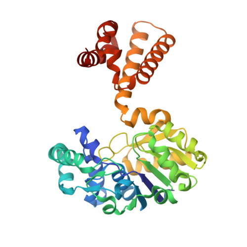
6YKA

Asymmetric [Fe]-hydrogenase from Methanolacinia paynteri apo and in complex with FeGP at 2.1-A resolution


Experimental Data Snapshot

  • Method: X-RAY DIFFRACTION
  • Resolution: 2.10 Å
  • R-Value Free: 0.235 
  • R-Value Work: 0.195 
  • R-Value Observed: 0.197 

Starting Model: experimental
View more details

wwPDB Validation   3D Report Full Report


Ligand Structure Quality Assessment 


This is version 1.1 of the entry. See complete history


Literature

The Hydride Transfer Process in NADP-dependent Methylene-tetrahydromethanopterin Dehydrogenase.

Huang, G.Wagner, T.Demmer, U.Warkentin, E.Ermler, U.Shima, S.

(2020) J Mol Biol 432: 2042-2054

  • DOI: https://doi.org/10.1016/j.jmb.2020.01.042
  • Primary Citation of Related Structures:  
    6TGE, 6TLK, 6TM3, 6YK9, 6YKA, 6YKB

  • PubMed Abstract: 

    NADP-dependent methylene-tetrahydromethanopterin (methylene-H 4 MPT) dehydrogenase (MtdA) catalyzes the reversible dehydrogenation of methylene-H 4 MPT to form methenyl-H 4 MPT + by using NADP + as a hydride acceptor. This hydride transfer reaction is involved in the oxidative metabolism from formaldehyde to CO 2 in methylotrophic and methanotrophic bacteria. Here, we report on the crystal structures of the ternary MtdA-substrate complexes from Methylorubrum extorquens AM1 obtained in open and closed forms. Their conversion is accomplished by opening/closing the active site cleft via a 15° rotation of the NADP, relative to the pterin domain. The 1.08 Å structure of the closed and active enzyme-NADP-methylene-H 4 MPT complex allows a detailed geometric analysis of the bulky substrates and a precise prediction of the hydride trajectory. Upon domain closure, the bulky substrate rings become compressed resulting in a tilt of the imidazolidine group of methylene-H 4 MPT that optimizes the geometry for hydride transfer. An additional 1.5 Å structure of MtdA in complex with the nonreactive NADP + and methenyl-H 4 MPT + revealed an extremely short distance between nicotinamide-C4 and imidazoline-C14a of 2.5 Å, which demonstrates the strong pressure imposed. The pterin-imidazolidine-phenyl butterfly angle of methylene-H 4 MPT bound to MtdA is smaller than that in the enzyme-free state but is similar to that in H 2 - and F 420 -dependent methylene-H 4 MPT dehydrogenases. The concept of compression-driven hydride transfer including quantum mechanical hydrogen tunneling effects, which are established for flavin- and NADP-dependent enzymes, can be expanded to hydride-transferring H 4 MPT-dependent enzymes.


  • Organizational Affiliation

    Max-Planck-Institut für terrestrische Mikrobiologie, Karl-von-Frisch Straße 10, 35043, Marburg, Germany.


Macromolecules
Find similar proteins by:  (by identity cutoff)  |  3D Structure
Entity ID: 1
MoleculeChains Sequence LengthOrganismDetailsImage
5,10-methenyltetrahydromethanopterin hydrogenase
A, B
342Methanolacinia paynteri G-2000Mutation(s): 0 
Gene Names: hmd
EC: 1.12.98.2
Entity Groups  
Sequence Clusters30% Identity50% Identity70% Identity90% Identity95% Identity100% Identity
Sequence Annotations
Expand
  • Reference Sequence
Small Molecules
Ligands 4 Unique
IDChains Name / Formula / InChI Key2D Diagram3D Interactions
FE9 (Subject of Investigation/LOI)
Query on FE9

Download Ideal Coordinates CCD File 
M [auth B]iron-guanylyl pyridinol cofactor
C21 H23 Fe N6 O13 P S
AEHOAZNVUAGELD-VPXBKTNXSA-K
PGE
Query on PGE

Download Ideal Coordinates CCD File 
G [auth A],
K [auth B]
TRIETHYLENE GLYCOL
C6 H14 O4
ZIBGPFATKBEMQZ-UHFFFAOYSA-N
GOL
Query on GOL

Download Ideal Coordinates CCD File 
I [auth A],
J [auth A],
N [auth B]
GLYCEROL
C3 H8 O3
PEDCQBHIVMGVHV-UHFFFAOYSA-N
EDO
Query on EDO

Download Ideal Coordinates CCD File 
C [auth A]
D [auth A]
E [auth A]
F [auth A]
H [auth A]
C [auth A],
D [auth A],
E [auth A],
F [auth A],
H [auth A],
L [auth B]
1,2-ETHANEDIOL
C2 H6 O2
LYCAIKOWRPUZTN-UHFFFAOYSA-N
Experimental Data & Validation

Experimental Data

  • Method: X-RAY DIFFRACTION
  • Resolution: 2.10 Å
  • R-Value Free: 0.235 
  • R-Value Work: 0.195 
  • R-Value Observed: 0.197 
  • Space Group: P 31 2 1
Unit Cell:
Length ( Å )Angle ( ˚ )
a = 79.481α = 90
b = 79.481β = 90
c = 179.337γ = 120
Software Package:
Software NamePurpose
SCALAdata scaling
BUSTERrefinement
PDB_EXTRACTdata extraction
XDSdata reduction
MOLREPphasing

Structure Validation

View Full Validation Report


Ligand Structure Quality Assessment 


Entry History & Funding Information

Deposition Data


Funding OrganizationLocationGrant Number
Max Planck SocietyGermany--
German Research Foundation (DFG)GermanyIron sulfur for life SH87/1-1
Ministry of Education (MoE, China)ChinaChina Scholarship Council

Revision History  (Full details and data files)

  • Version 1.0: 2021-02-17
    Type: Initial release
  • Version 1.1: 2024-01-24
    Changes: Data collection, Database references, Refinement description