6ZFP

Cryo-EM structure of DNA-PKcs (State 2)


Experimental Data Snapshot

  • Method: ELECTRON MICROSCOPY
  • Resolution: 3.24 Å
  • Aggregation State: PARTICLE 
  • Reconstruction Method: SINGLE PARTICLE 

wwPDB Validation   3D Report Full Report


This is version 1.2 of the entry. See complete history


Literature

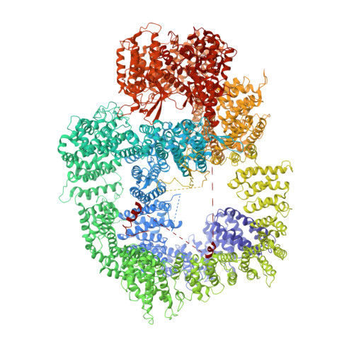
Dimers of DNA-PK create a stage for DNA double-strand break repair.

Chaplin, A.K.Hardwick, S.W.Liang, S.Kefala Stavridi, A.Hnizda, A.Cooper, L.R.De Oliveira, T.M.Chirgadze, D.Y.Blundell, T.L.

(2021) Nat Struct Mol Biol 28: 13-19

  • DOI: https://doi.org/10.1038/s41594-020-00517-x
  • Primary Citation of Related Structures:  
    6ZFP, 6ZH2, 6ZH4, 6ZH6, 6ZH8, 6ZHA, 6ZHE

  • PubMed Abstract: 

    DNA double-strand breaks are the most dangerous type of DNA damage and, if not repaired correctly, can lead to cancer. In humans, Ku70/80 recognizes DNA broken ends and recruits the DNA-dependent protein kinase catalytic subunit (DNA-PKcs) to form DNA-dependent protein kinase holoenzyme (DNA-PK) in the process of non-homologous end joining (NHEJ). We present a 2.8-Å-resolution cryo-EM structure of DNA-PKcs, allowing precise amino acid sequence registration in regions uninterpreted in previous 4.3-Å X-ray maps. We also report a cryo-EM structure of DNA-PK at 3.5-Å resolution and reveal a dimer mediated by the Ku80 C terminus. Central to dimer formation is a domain swap of the conserved C-terminal helix of Ku80. Our results suggest a new mechanism for NHEJ utilizing a DNA-PK dimer to bring broken DNA ends together. Furthermore, drug inhibition of NHEJ in combination with chemo- and radiotherapy has proved successful, making these models central to structure-based drug targeting efforts.


  • Organizational Affiliation

    Department of Biochemistry, University of Cambridge, Cambridge, UK.


Macromolecules
Find similar proteins by:  (by identity cutoff)  |  3D Structure
Entity ID: 1
MoleculeChains Sequence LengthOrganismDetailsImage
DNA-dependent protein kinase catalytic subunit,DNA-PKcs,DNA-PKcs4,156Homo sapiensMutation(s): 0 
Gene Names: PRKDCHYRCHYRC1
EC: 2.7.11.1
UniProt & NIH Common Fund Data Resources
Find proteins for P78527 (Homo sapiens)
Explore P78527 
Go to UniProtKB:  P78527
PHAROS:  P78527
GTEx:  ENSG00000253729 
Entity Groups  
Sequence Clusters30% Identity50% Identity70% Identity90% Identity95% Identity100% Identity
UniProt GroupP78527
Sequence Annotations
Expand
  • Reference Sequence
Experimental Data & Validation

Experimental Data

  • Method: ELECTRON MICROSCOPY
  • Resolution: 3.24 Å
  • Aggregation State: PARTICLE 
  • Reconstruction Method: SINGLE PARTICLE 
EM Software:
TaskSoftware PackageVersion
RECONSTRUCTIONcryoSPARC

Structure Validation

View Full Validation Report


Entry History & Funding Information

Deposition Data


Funding OrganizationLocationGrant Number
Wellcome TrustUnited Kingdom200814/Z/16/Z

Revision History  (Full details and data files)

  • Version 1.0: 2020-10-21
    Type: Initial release
  • Version 1.1: 2020-11-04
    Changes: Database references
  • Version 1.2: 2021-01-20
    Changes: Database references