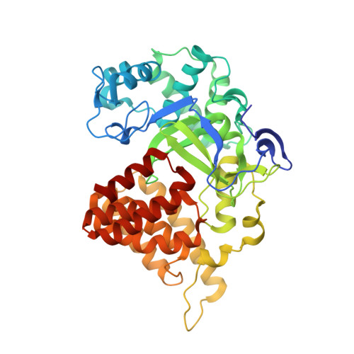
6CBY | pdb_00006cby

Crystal structure of human SET and MYND Domain Containing protein 2 with MTF9975


Experimental Data Snapshot

  • Method: X-RAY DIFFRACTION
  • Resolution: 2.55 Å
  • R-Value Free: 
    0.258 (Depositor), 0.262 (DCC) 
  • R-Value Work: 
    0.199 (Depositor), 0.209 (DCC) 
  • R-Value Observed: 
    0.201 (Depositor) 

wwPDB Validation 3D Report Full Report

Validation slider image for 6CBY

Ligand Structure Quality Assessment 


This is version 1.1 of the entry. See complete history. 

Literature

Selective, Small-Molecule Co-Factor Binding Site Inhibition of a Su(var)3-9, Enhancer of Zeste, Trithorax Domain Containing Lysine Methyltransferase.

Taylor, A.P., Swewczyk, M., Kennedy, S., Trush, V.V., Wu, H., Zeng, H., Dong, A., Ferreira de Freitas, R., Tatlock, J., Kumpf, R.A., Wythes, M., Casimiro-Garcia, A., Denny, R.A., Parikh, M.D., Li, F., Barsyte-Lovejoy, D., Schapira, M., Vedadi, M., Brown, P.J., Arrowsmith, C.H., Owen, D.R.

(2019) J Med Chem 62: 7669-7683

  • DOI: https://doi.org/10.1021/acs.jmedchem.9b00112
  • Primary Citation Related Structures: 
    6CBY

  • PubMed Abstract: 

    The first chemical probe to primarily occupy the co-factor binding site of a Su(var)3-9, enhancer of a zeste, trithorax (SET) domain containing protein lysine methyltransferase (PKMT) is reported. Protein methyltransferases require S -adenosylmethionine (SAM) as a co-factor (methyl donor) for enzymatic activity. However, SAM itself represents a poor medicinal chemistry starting point for a selective, cell-active inhibitor given its extreme physicochemical properties and its role in multiple cellular processes. A previously untested medicinal chemistry strategy of deliberate file enrichment around molecules bearing the hallmarks of SAM, but with improved lead-like properties from the outset, yielded viable hits against SET and MYND domain-containing protein 2 (SMYD2) that were shown to bind in the co-factor site. These leads were optimized to identify a highly biochemically potent, PKMT-selective, and cell-active chemical probe. While substrate-based inhibitors of PKMTs are known, this represents a novel, co-factor-derived strategy for the inhibition of SMYD2 which may also prove applicable to lysine methyltransferase family members previously thought of as intractable.


  • Organizational Affiliation: 
    • Pfizer Medicine Design, Medicinal Chemistry , Pfizer Worldwide Research and Development , Eastern Point Road , Groton , Connecticut 06340 , United States.

Macromolecule Content 

  • Total Structure Weight: 101.17 kDa 
  • Atom Count: 6,540 
  • Modeled Residue Count: 850 
  • Deposited Residue Count: 868 
  • Unique protein chains: 1

Macromolecules

Find similar proteins by:|  3D Structure
Entity ID: 1
MoleculeChains  Sequence LengthOrganismDetailsImage
N-lysine methyltransferase SMYD2
A, B
434Homo sapiensMutation(s): 0 
Gene Names: SMYD2, KMT3C
EC: 2.1.1 (PDB Primary Data), 2.1.1.43 (PDB Primary Data), 2.1.1.354 (UniProt)
UniProt & NIH Common Fund Data Resources
Find proteins for Q9NRG4 (Homo sapiens)
Explore Q9NRG4 
Go to UniProtKB:  Q9NRG4
PHAROS:  Q9NRG4
GTEx:  ENSG00000143499 
Entity Groups
Sequence Clusters30% Identity50% Identity70% Identity90% Identity95% Identity100% Identity
UniProt GroupQ9NRG4
Sequence Annotations
Expand
Reference Sequence

Small Molecules

Ligands 2 Unique
IDChains Name / Formula / InChI Key2D Diagram3D Interactions
EW4

Query on EW4


Download:Ideal Coordinates CCD File
F [auth A],
J [auth B]
[3-(4-amino-6-methyl-1H-imidazo[4,5-c]pyridin-1-yl)-3-methylazetidin-1-yl][1-({1-[(1R)-cyclohept-2-en-1-yl]piperidin-4-yl}methyl)-1H-pyrrol-3-yl]methanone
C29 H39 N7 O
AVKAVKZKOFSSMY-DEOSSOPVSA-N
ZN

Query on ZN


Download:Ideal Coordinates CCD File
C [auth A]
D [auth A]
E [auth A]
G [auth B]
H [auth B]
C [auth A],
D [auth A],
E [auth A],
G [auth B],
H [auth B],
I [auth B]
ZINC ION
Zn
PTFCDOFLOPIGGS-UHFFFAOYSA-N
Binding Affinity Annotations 
IDSourceBinding Affinity
EW4 BindingDB:  6CBY IC50: min: 8, max: 1300 (nM) from 3 assay(s)

Experimental Data & Validation

Experimental Data

  • Method: X-RAY DIFFRACTION
  • Resolution: 2.55 Å
  • R-Value Free:  0.258 (Depositor), 0.262 (DCC) 
  • R-Value Work:  0.199 (Depositor), 0.209 (DCC) 
  • R-Value Observed: 0.201 (Depositor) 
Space Group: P 1 21 1
Unit Cell:
Length ( Å )Angle ( ˚ )
a = 57.819α = 90
b = 117.579β = 92.42
c = 65.013γ = 90
Software Package:
Software NamePurpose
BUSTERrefinement
SCALEPACKdata scaling
PDB_EXTRACTdata extraction
HKL-3000data reduction
PHASERphasing

Structure Validation

View Full Validation Report


Ligand Structure Quality Assessment 


Entry History 

Revision History  (Full details and data files)

  • Version 1.0: 2018-03-14
    Type: Initial release
  • Version 1.1: 2023-12-27
    Changes: Data collection, Database references