Human beta-defensin 2 killsCandida albicansthrough phosphatidylinositol 4,5-bisphosphate-mediated membrane permeabilization.
Jarva, M., Phan, T.K., Lay, F.T., Caria, S., Kvansakul, M., Hulett, M.D.(2018) Sci Adv 4: eaat0979-eaat0979
- PubMed: 30050988 Search on PubMedSearch on PubMed Central
- DOI: https://doi.org/10.1126/sciadv.aat0979
- Primary Citation Related Structures:
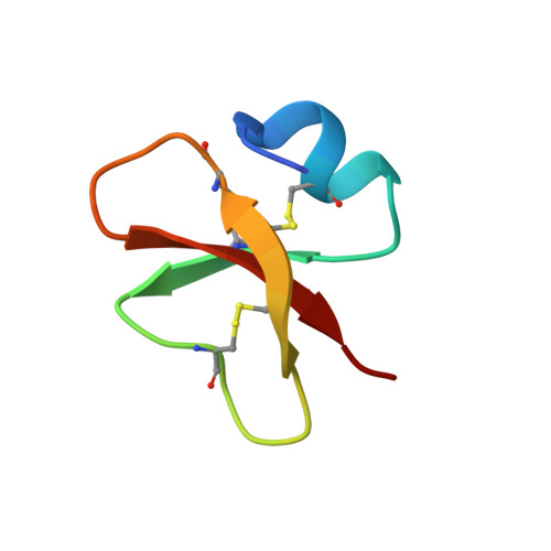
6CS9 - PubMed Abstract:
Human defensins belong to a subfamily of the cationic antimicrobial peptides and act as a first line of defense against invading microbes. Their often broad-spectrum antimicrobial and antitumor activities make them attractive for therapeutic development; however, their precise molecular mechanism(s) of action remains to be defined. We show that human β-defensin 2 (HBD-2) permeabilizes Candida albicans cell membranes via a mechanism targeting the plasma membrane lipid phosphatidylinositol 4,5-bisphosphate (PIP 2 ). We determined the structure of HBD-2 bound to PIP 2 , which revealed two distinct PIP 2 -binding sites, and showed, using functional assays, that mutations in these sites ablate PIP 2 -mediated fungal growth inhibition by HBD-2. Our study provides the first insight into lipid-mediated human defensin membrane permeabilization at an atomic level and reveals a unique mode of lipid engagement to permeabilize cell membranes.
- Department of Biochemistry and Genetics, La Trobe Institute for Molecular Science, La Trobe University, Melbourne, Australia.
Organizational Affiliation: