6V0P | pdb_00006v0p

PRMT5 complex bound to covalent PBM inhibitor BRD6711


Experimental Data Snapshot

  • Method: X-RAY DIFFRACTION
  • Resolution: 1.88 Å
  • R-Value Free: 
    0.230 (Depositor), 0.234 (DCC) 
  • R-Value Work: 
    0.187 (Depositor), 0.193 (DCC) 

Starting Model: experimental
View more details

wwPDB Validation 3D Report Full Report

Validation slider image for 6V0P

Ligand Structure Quality Assessment 


This is version 1.3 of the entry. See complete history

Literature

Discovery of a First-in-Class Inhibitor of the PRMT5-Substrate Adaptor Interaction.

McKinney, D.C.McMillan, B.J.Ranaghan, M.J.Moroco, J.A.Brousseau, M.Mullin-Bernstein, Z.O'Keefe, M.McCarren, P.Mesleh, M.F.Mulvaney, K.M.Robinson, F.Singh, R.Bajrami, B.Wagner, F.F.Hilgraf, R.Drysdale, M.J.Campbell, A.J.Skepner, A.Timm, D.E.Porter, D.Kaushik, V.K.Sellers, W.R.Ianari, A.

(2021) J Med Chem 64: 11148-11168

  • DOI: https://doi.org/10.1021/acs.jmedchem.1c00507
  • Primary Citation Related Structures: 
    6V0P, 7M05

  • PubMed Abstract: 

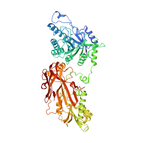
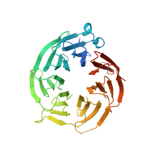
    PRMT5 and its substrate adaptor proteins (SAPs), pICln and Riok1, are synthetic lethal dependencies in MTAP-deleted cancer cells. SAPs share a conserved PRMT5 binding motif (PBM) which mediates binding to a surface of PRMT5 distal to the catalytic site. This interaction is required for methylation of several PRMT5 substrates, including histone and spliceosome complexes. We screened for small molecule inhibitors of the PRMT5-PBM interaction and validated a compound series which binds to the PRMT5-PBM interface and directly inhibits binding of SAPs. Mode of action studies revealed the formation of a covalent bond between a halogenated pyridazinone group and cysteine 278 of PRMT5. Optimization of the starting hit produced a lead compound, BRD0639, which engages the target in cells, disrupts PRMT5-RIOK1 complexes, and reduces substrate methylation. BRD0639 is a first-in-class PBM-competitive inhibitor that can support studies of PBM-dependent PRMT5 activities and the development of novel PRMT5 inhibitors that selectively target these functions.


  • Organizational Affiliation
    • Center for the Development of Therapeutics, The Broad Institute of MIT and Harvard, 415 Main Street, Cambridge, Massachusetts 02142, United States.

Macromolecule Content 

  • Total Structure Weight: 110.48 kDa 
  • Atom Count: 7,987 
  • Modeled Residue Count: 916 
  • Deposited Residue Count: 979 
  • Unique protein chains: 2

Macromolecules

Find similar proteins by:|  3D Structure
Entity ID: 1
MoleculeChains  Sequence LengthOrganismDetailsImage
Protein arginine N-methyltransferase 5637Homo sapiensMutation(s): 0 
Gene Names: PRMT5HRMT1L5IBP72JBP1SKB1
EC: 2.1.1.320
UniProt & NIH Common Fund Data Resources
Find proteins for O14744 (Homo sapiens)
Explore O14744 
Go to UniProtKB:  O14744
PHAROS:  O14744
GTEx:  ENSG00000100462 
Entity Groups
Sequence Clusters30% Identity50% Identity70% Identity90% Identity95% Identity100% Identity
UniProt GroupO14744
Sequence Annotations
Expand
Reference Sequence
Find similar proteins by:|  3D Structure
Entity ID: 2
MoleculeChains  Sequence LengthOrganismDetailsImage
Methylosome protein 50342Homo sapiensMutation(s): 0 
Gene Names: WDR77MEP50WD45HKMT1069Nbla10071
UniProt & NIH Common Fund Data Resources
Find proteins for Q9BQA1 (Homo sapiens)
Explore Q9BQA1 
Go to UniProtKB:  Q9BQA1
PHAROS:  Q9BQA1
GTEx:  ENSG00000116455 
Entity Groups
Sequence Clusters30% Identity50% Identity70% Identity90% Identity95% Identity100% Identity
UniProt GroupQ9BQA1
Sequence Annotations
Expand
Reference Sequence

Small Molecules

Ligands 4 Unique
IDChains Name / Formula / InChI Key2D Diagram3D Interactions
SFG

Query on SFG


Download:Ideal Coordinates CCD File
F [auth A]SINEFUNGIN
C15 H23 N7 O5
LMXOHSDXUQEUSF-YECHIGJVSA-N
QN4
(Subject of Investigation/LOI)

Query on QN4


Download:Ideal Coordinates CCD File
C [auth A]2-(5-chloro-6-oxopyridazin-1(6H)-yl)-N-(4-methyl-3-sulfamoylphenyl)acetamide
C13 H13 Cl N4 O4 S
SJCYOELGOMZSNG-UHFFFAOYSA-N
GOL

Query on GOL


Download:Ideal Coordinates CCD File
G [auth A],
H [auth A]
GLYCEROL
C3 H8 O3
PEDCQBHIVMGVHV-UHFFFAOYSA-N
CL

Query on CL


Download:Ideal Coordinates CCD File
D [auth A],
E [auth A]
CHLORIDE ION
Cl
VEXZGXHMUGYJMC-UHFFFAOYSA-M

Experimental Data & Validation

Experimental Data

  • Method: X-RAY DIFFRACTION
  • Resolution: 1.88 Å
  • R-Value Free:  0.230 (Depositor), 0.234 (DCC) 
  • R-Value Work:  0.187 (Depositor), 0.193 (DCC) 
Space Group: I 2 2 2
Unit Cell:
Length ( Å )Angle ( ˚ )
a = 99.11α = 90
b = 138.57β = 90
c = 178.43γ = 90
Software Package:
Software NamePurpose
XDSdata reduction
BUSTERrefinement
PDB_EXTRACTdata extraction
STARANISOdata scaling
PHASERphasing

Structure Validation

View Full Validation Report


Ligand Structure Quality Assessment 


Entry History 

Deposition Data

Revision History  (Full details and data files)

  • Version 1.0: 2020-11-25
    Type: Initial release
  • Version 1.1: 2021-12-08
    Changes: Advisory, Database references
  • Version 1.2: 2023-10-11
    Changes: Data collection, Refinement description
  • Version 1.3: 2024-10-23
    Changes: Structure summary