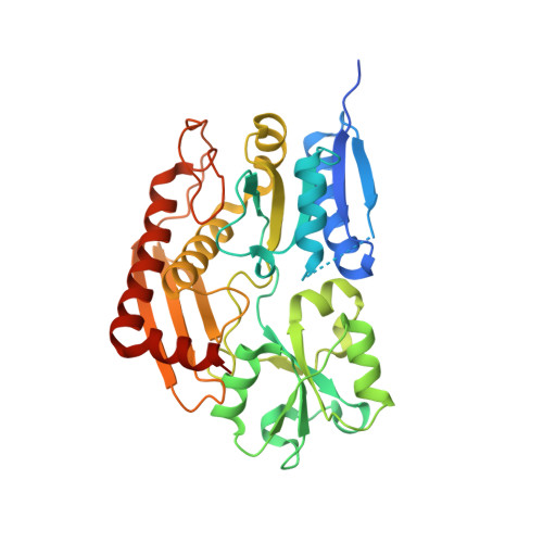
7AAJ

Human porphobilinogen deaminase in complex with cofactor


Experimental Data Snapshot

  • Method: X-RAY DIFFRACTION
  • Resolution: 1.80 Å
  • R-Value Free: 0.294 
  • R-Value Work: 0.249 
  • R-Value Observed: 0.251 

Starting Model: experimental
View more details

wwPDB Validation   3D Report Full Report


Ligand Structure Quality Assessment 


This is version 1.2 of the entry. See complete history


Literature

Characterization of porphobilinogen deaminase mutants reveals that arginine-173 is crucial for polypyrrole elongation mechanism.

Bustad, H.J.Kallio, J.P.Laitaoja, M.Toska, K.Kursula, I.Martinez, A.Janis, J.

(2021) iScience 24: 102152-102152

  • DOI: https://doi.org/10.1016/j.isci.2021.102152
  • Primary Citation of Related Structures:  
    7AAJ, 7AAK

  • PubMed Abstract: 

    Porphobilinogen deaminase (PBGD), the third enzyme in the heme biosynthesis, catalyzes the sequential coupling of four porphobilinogen (PBG) molecules into a heme precursor. Mutations in PBGD are associated with acute intermittent porphyria (AIP), a rare metabolic disorder. We used Fourier transform ion cyclotron resonance mass spectrometry (FT-ICR MS) to demonstrate that wild-type PBGD and AIP-associated mutant R167W both existed as holoenzymes (E holo ) covalently attached to the dipyrromethane cofactor, and three intermediate complexes, ES, ES 2 , and ES 3 , where S represents PBG. In contrast, only ES 2 was detected in AIP-associated mutant R173W, indicating that the formation of ES 3 is inhibited. The R173W crystal structure in the ES 2 -state revealed major rearrangements of the loops around the active site, compared to wild-type PBGD in the E holo -state. These results contribute to elucidating the structural pathogenesis of two common AIP-associated mutations and reveal the important structural role of Arg173 in the polypyrrole elongation mechanism.


  • Organizational Affiliation

    Department of Biomedicine, University of Bergen, Jonas Lies vei 91, 5009 Bergen, Norway.


Macromolecules
Find similar proteins by:  (by identity cutoff)  |  3D Structure
Entity ID: 1
MoleculeChains Sequence LengthOrganismDetailsImage
Porphobilinogen deaminaseA [auth B],
B [auth A]
363Homo sapiensMutation(s): 0 
Gene Names: HMBSPBGDUPS
EC: 2.5.1.61
UniProt & NIH Common Fund Data Resources
Find proteins for P08397 (Homo sapiens)
Explore P08397 
Go to UniProtKB:  P08397
PHAROS:  P08397
GTEx:  ENSG00000256269 
Entity Groups  
Sequence Clusters30% Identity50% Identity70% Identity90% Identity95% Identity100% Identity
UniProt GroupP08397
Sequence Annotations
Expand
  • Reference Sequence
Experimental Data & Validation

Experimental Data

  • Method: X-RAY DIFFRACTION
  • Resolution: 1.80 Å
  • R-Value Free: 0.294 
  • R-Value Work: 0.249 
  • R-Value Observed: 0.251 
  • Space Group: P 21 21 21
Unit Cell:
Length ( Å )Angle ( ˚ )
a = 81.163α = 90
b = 84.608β = 90
c = 108.913γ = 90
Software Package:
Software NamePurpose
XDSdata processing
XDSdata reduction
Aimlessdata scaling
PHASERphasing
PHENIXrefinement
Cootmodel building

Structure Validation

View Full Validation Report


Ligand Structure Quality Assessment 


Entry History 

Deposition Data

Revision History  (Full details and data files)

  • Version 1.0: 2021-02-17
    Type: Initial release
  • Version 1.1: 2021-03-17
    Changes: Database references
  • Version 1.2: 2024-01-31
    Changes: Data collection, Database references, Refinement description