7AEW

14-3-3 sigma bound to bis-phosphorylated aminopeptidase N (APN, CD13) via canonical and non-canonical binding motifs


Experimental Data Snapshot

  • Method: X-RAY DIFFRACTION
  • Resolution: 1.20 Å
  • R-Value Free: 0.184 
  • R-Value Work: 0.162 

wwPDB Validation   3D Report Full Report


This is version 1.4 of the entry. See complete history


Literature

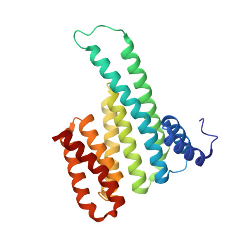
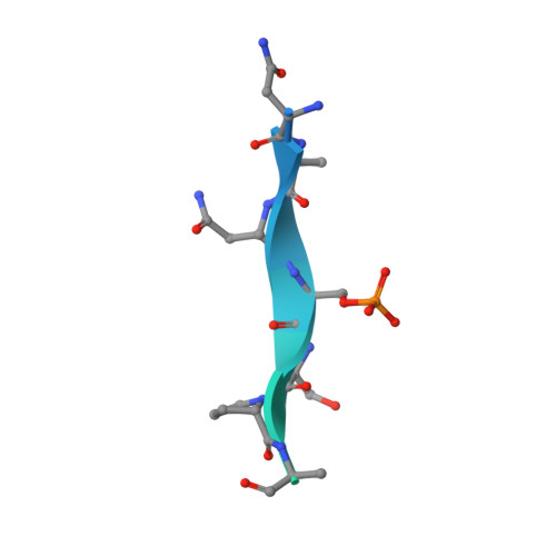
MMP activation-associated aminopeptidase N reveals a bivalent 14-3-3 binding motif.

Kiehstaller, S.Ottmann, C.Hennig, S.

(2020) J Biol Chem 295: 18266-18275

  • DOI: https://doi.org/10.1074/jbc.RA120.014708
  • Primary Citation of Related Structures:  
    6XWD, 7AEW

  • PubMed Abstract: 

    Aminopeptidase N (APN, CD13) is a transmembrane ectopeptidase involved in many crucial cellular functions. Besides its role as a peptidase, APN also mediates signal transduction and is involved in the activation of matrix metalloproteinases (MMPs). MMPs function in tissue remodeling within the extracellular space and are therefore involved in many human diseases, such as fibrosis, rheumatoid arthritis, tumor angiogenesis, and metastasis, as well as viral infections. However, the exact mechanism that leads to APN-driven MMP activation is unclear. It was previously shown that extracellular 14-3-3 adapter proteins bind to APN and thereby induce the transcription of MMPs. As a first step, we sought to identify potential 14-3-3-binding sites in the APN sequence. We constructed a set of phosphorylated peptides derived from APN to probe for interactions. We identified and characterized a canonical 14-3-3-binding site ( site 1 ) within the flexible, structurally unresolved N-terminal APN region using direct binding fluorescence polarization assays and thermodynamic analysis. In addition, we identified a secondary, noncanonical binding site ( site 2 ), which enhances the binding affinity in combination with site 1 by many orders of magnitude. Finally, we solved crystal structures of 14-3-3σ bound to mono- and bis-phosphorylated APN-derived peptides, which revealed atomic details of the binding mode of mono- and bivalent 14-3-3 interactions. Therefore, our findings shed some light on the first steps of APN-mediated MMP activation and open the field for further investigation of this important signaling pathway.


  • Organizational Affiliation

    Department of Chemistry and Pharmaceutical Sciences, VU University Amsterdam, Amsterdam, Netherlands; Amsterdam Institute of Molecular and Life Sciences (AIMMS), VU University Amsterdam, Amsterdam, Netherlands.


Macromolecules
Find similar proteins by:  (by identity cutoff)  |  3D Structure
Entity ID: 1
MoleculeChains Sequence LengthOrganismDetailsImage
14-3-3 protein sigmaA [auth AAA]236Homo sapiensMutation(s): 0 
Gene Names: SFNHME1
UniProt & NIH Common Fund Data Resources
Find proteins for P31947 (Homo sapiens)
Explore P31947 
Go to UniProtKB:  P31947
PHAROS:  P31947
GTEx:  ENSG00000175793 
Entity Groups  
Sequence Clusters30% Identity50% Identity70% Identity90% Identity95% Identity100% Identity
UniProt GroupP31947
Sequence Annotations
Expand
  • Reference Sequence
Find similar proteins by:  (by identity cutoff)  |  3D Structure
Entity ID: 2
MoleculeChains Sequence LengthOrganismDetailsImage
Aminopeptidase NB [auth CCC],
C [auth BBB]
38Homo sapiensMutation(s): 0 
EC: 3.4.11.2
UniProt & NIH Common Fund Data Resources
Find proteins for P15144 (Homo sapiens)
Explore P15144 
Go to UniProtKB:  P15144
PHAROS:  P15144
GTEx:  ENSG00000166825 
Entity Groups  
Sequence Clusters30% Identity50% Identity70% Identity90% Identity95% Identity100% Identity
UniProt GroupP15144
Sequence Annotations
Expand
  • Reference Sequence
Experimental Data & Validation

Experimental Data

  • Method: X-RAY DIFFRACTION
  • Resolution: 1.20 Å
  • R-Value Free: 0.184 
  • R-Value Work: 0.162 
  • Space Group: C 2 2 21
Unit Cell:
Length ( Å )Angle ( ˚ )
a = 82.21α = 90
b = 112.06β = 90
c = 62.61γ = 90
Software Package:
Software NamePurpose
XDSdata reduction
XSCALEdata scaling
PHASERphasing
REFMACrefinement

Structure Validation

View Full Validation Report


Entry History 

Deposition Data

Revision History  (Full details and data files)

  • Version 1.0: 2020-11-04
    Type: Initial release
  • Version 1.1: 2020-11-11
    Changes: Database references
  • Version 1.2: 2021-01-06
    Changes: Database references
  • Version 1.3: 2022-06-08
    Changes: Database references, Derived calculations
  • Version 1.4: 2024-01-31
    Changes: Data collection, Derived calculations, Refinement description