Getting a Grip on the Undrugged: Targeting beta-Catenin with Fragment-Based Methods.
Kessler, D., Mayer, M., Zahn, S.K., Zeeb, M., Wohrle, S., Bergner, A., Bruchhaus, J., Ciftci, T., Dahmann, G., Dettling, M., Dobel, S., Fuchs, J.E., Geist, L., Hela, W., Kofink, C., Kousek, R., Moser, F., Puchner, T., Rumpel, K., Scharnweber, M., Werni, P., Wolkerstorfer, B., Breitsprecher, D., Baaske, P., Pearson, M., McConnell, D.B., Bottcher, J.(2021) ChemMedChem 16: 1420-1424
- PubMed: 33275320 Search on PubMedSearch on PubMed Central
- DOI: https://doi.org/10.1002/cmdc.202000839
- Primary Citation Related Structures:
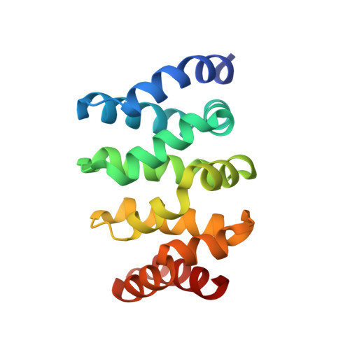
7AFW - PubMed Abstract:
Aberrant WNT pathway activation, leading to nuclear accumulation of β-catenin, is a key oncogenic driver event. Mutations in the tumor suppressor gene APC lead to impaired proteasomal degradation of β-catenin and subsequent nuclear translocation. Restoring cellular degradation of β-catenin represents a potential therapeutic strategy. Here, we report the fragment-based discovery of a small molecule binder to β-catenin, including the structural elucidation of the binding mode by X-ray crystallography. The difficulty in drugging β-catenin was confirmed as the primary screening campaigns identified only few and very weak hits. Iterative virtual and NMR screening techniques were required to discover a compound with sufficient potency to be able to obtain an X-ray co-crystal structure. The binding site is located between armadillo repeats two and three, adjacent to the BCL9 and TCF4 binding sites. Genetic studies show that it is unlikely to be useful for the development of protein-protein interaction inhibitors but structural information and established assays provide a solid basis for a prospective optimization towards β-catenin proteolysis targeting chimeras (PROTACs) as alternative modality.
- Boehringer Ingelheim RCV GmbH & Co KG, Dr.-Boehringer-Gasse 5-11, 1121, Vienna, Austria.
Organizational Affiliation: