7B2L

Structure of the endocytic adaptor complex AENTH


Experimental Data Snapshot

  • Method: ELECTRON MICROSCOPY
  • Resolution: 3.90 Å
  • Aggregation State: PARTICLE 
  • Reconstruction Method: SINGLE PARTICLE 

wwPDB Validation   3D Report Full Report


This is version 1.1 of the entry. See complete history


Literature

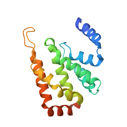
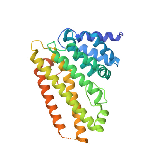
Structure of the endocytic adaptor complex reveals the basis for efficient membrane anchoring during clathrin-mediated endocytosis.

Lizarrondo, J.Klebl, D.P.Niebling, S.Abella, M.Schroer, M.A.Mertens, H.D.T.Veith, K.Thuenauer, R.Svergun, D.I.Skruzny, M.Sobott, F.Muench, S.P.Garcia-Alai, M.M.

(2021) Nat Commun 12: 2889-2889

  • DOI: https://doi.org/10.1038/s41467-021-23151-7
  • Primary Citation of Related Structures:  
    7B2L

  • PubMed Abstract: 

    During clathrin-mediated endocytosis, a complex and dynamic network of protein-membrane interactions cooperate to achieve membrane invagination. Throughout this process in yeast, endocytic coat adaptors, Sla2 and Ent1, must remain attached to the plasma membrane to transmit force from the actin cytoskeleton required for successful membrane invagination. Here, we present a cryo-EM structure of a 16-mer complex of the ANTH and ENTH membrane-binding domains from Sla2 and Ent1 bound to PIP 2 that constitutes the anchor to the plasma membrane. Detailed in vitro and in vivo mutagenesis of the complex interfaces delineate the key interactions for complex formation and deficient cell growth phenotypes demonstrate its biological relevance. A hetero-tetrameric unit binds PIP 2 molecules at the ANTH-ENTH interfaces and can form larger assemblies to contribute to membrane remodeling. Finally, a time-resolved small-angle X-ray scattering study of the interaction of these adaptor domains in vitro suggests that ANTH and ENTH domains have evolved to achieve a fast subsecond timescale assembly in the presence of PIP 2 and do not require further proteins to form a stable complex. Together, these findings provide a molecular understanding of an essential piece in the molecular puzzle of clathrin-coated endocytic sites.


  • Organizational Affiliation

    European Molecular Biology Laboratory, Hamburg Outstation, Hamburg, Germany.


Macromolecules
Find similar proteins by:  (by identity cutoff)  |  3D Structure
Entity ID: 1
MoleculeChains Sequence LengthOrganismDetailsImage
ENTH domain of epsin Ent1162Saccharomyces cerevisiae S288CMutation(s): 0 
Gene Names: ENT1YDL161W
UniProt
Find proteins for Q12518 (Saccharomyces cerevisiae (strain ATCC 204508 / S288c))
Explore Q12518 
Go to UniProtKB:  Q12518
Entity Groups  
Sequence Clusters30% Identity50% Identity70% Identity90% Identity95% Identity100% Identity
UniProt GroupQ12518
Sequence Annotations
Expand
  • Reference Sequence
Find similar proteins by:  (by identity cutoff)  |  3D Structure
Entity ID: 2
MoleculeChains Sequence LengthOrganismDetailsImage
ANTH domain of Sla2291Saccharomyces cerevisiae S288CMutation(s): 0 
Gene Names: SLA2END4MOP2UFG1YNL243WN1102
UniProt
Find proteins for P33338 (Saccharomyces cerevisiae (strain ATCC 204508 / S288c))
Explore P33338 
Go to UniProtKB:  P33338
Entity Groups  
Sequence Clusters30% Identity50% Identity70% Identity90% Identity95% Identity100% Identity
UniProt GroupP33338
Sequence Annotations
Expand
  • Reference Sequence
Small Molecules
Ligands 1 Unique
IDChains Name / Formula / InChI Key2D Diagram3D Interactions
PIO (Subject of Investigation/LOI)
Query on PIO

Download Ideal Coordinates CCD File 
AA [auth K]
BA [auth K]
CA [auth L]
DA [auth M]
EA [auth M]
AA [auth K],
BA [auth K],
CA [auth L],
DA [auth M],
EA [auth M],
FA [auth P],
GA [auth P],
HA [auth Q],
IA [auth R],
JA [auth R],
Q [auth A],
R [auth A],
S [auth B],
T [auth C],
U [auth C],
V [auth F],
W [auth F],
X [auth G],
Y [auth H],
Z [auth H]
[(2R)-2-octanoyloxy-3-[oxidanyl-[(1R,2R,3S,4R,5R,6S)-2,3,6-tris(oxidanyl)-4,5-diphosphonooxy-cyclohexyl]oxy-phosphoryl]oxy-propyl] octanoate
C25 H49 O19 P3
XLNCEHRXXWQMPK-MJUMVPIBSA-N
Experimental Data & Validation

Experimental Data

  • Method: ELECTRON MICROSCOPY
  • Resolution: 3.90 Å
  • Aggregation State: PARTICLE 
  • Reconstruction Method: SINGLE PARTICLE 
EM Software:
TaskSoftware PackageVersion
MODEL REFINEMENTISOLDE
MODEL REFINEMENTPHENIX

Structure Validation

View Full Validation Report


Entry History & Funding Information

Deposition Data


Funding OrganizationLocationGrant Number
German Research Foundation (DFG)GermanySK 305/1-1
Wellcome TrustUnited Kingdom108466/Z/15/Z

Revision History  (Full details and data files)

  • Version 1.0: 2021-05-05
    Type: Initial release
  • Version 1.1: 2021-06-02
    Changes: Database references