7B5Q

Cryo-EM structure of the human CAK bound to ICEC0942 (PHENIX-OPLS3e)


Experimental Data Snapshot

  • Method: ELECTRON MICROSCOPY
  • Resolution: 2.50 Å
  • Aggregation State: PARTICLE 
  • Reconstruction Method: SINGLE PARTICLE 

wwPDB Validation   3D Report Full Report


This is version 1.2 of the entry. See complete history


Literature

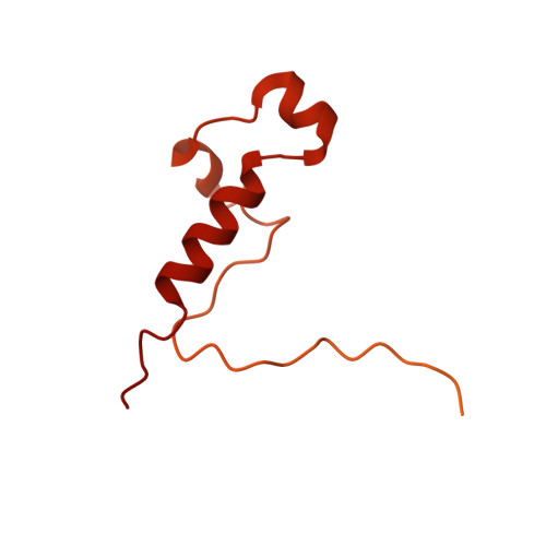
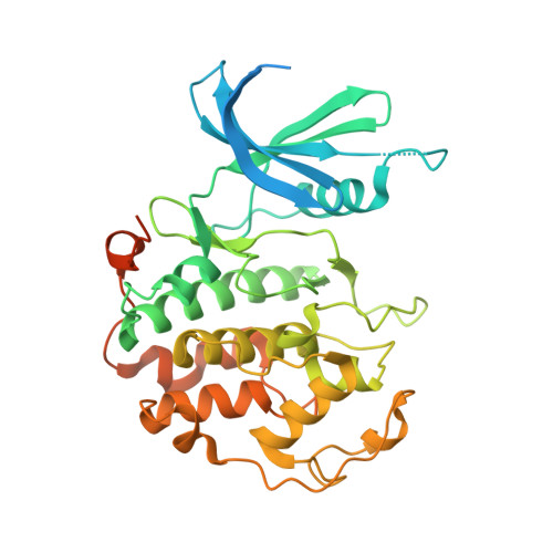
2.5 angstrom -resolution structure of human CDK-activating kinase bound to the clinical inhibitor ICEC0942.

Greber, B.J.Remis, J.Ali, S.Nogales, E.

(2021) Biophys J 120: 677-686

  • DOI: https://doi.org/10.1016/j.bpj.2020.12.030
  • Primary Citation of Related Structures:  
    7B5O, 7B5Q

  • PubMed Abstract: 

    The human CDK-activating kinase (CAK), composed of CDK7, cyclin H, and MAT1, is involved in the control of transcription initiation and the cell cycle. Because of these activities, it has been identified as a promising target for cancer chemotherapy. A number of CDK7 inhibitors have entered clinical trials, among them ICEC0942 (also known as CT7001). Structural information can aid in improving the affinity and specificity of such drugs or drug candidates, reducing side effects in patients. Here, we have determined the structure of the human CAK in complex with ICEC0942 at 2.5 Å-resolution using cryogenic electron microscopy. Our structure reveals conformational differences of ICEC0942 compared with previous X-ray crystal structures of the CDK2-bound complex, and highlights the critical ability of cryogenic electron microscopy to resolve structures of drug-bound protein complexes without the need to crystalize the protein target.


  • Organizational Affiliation

    Division of Structural Biology, The Institute of Cancer Research, London, United Kingdom; California Institute for Quantitative Biosciences (QB3), University of California, Berkeley, Berkeley, California; Molecular Biophysics and Integrative Bio-Imaging Division, Lawrence Berkeley National Laboratory, Berkeley, California. Electronic address: basil.greber@icr.ac.uk.


Macromolecules
Find similar proteins by:  (by identity cutoff)  |  3D Structure
Entity ID: 1
MoleculeChains Sequence LengthOrganismDetailsImage
CDK-activating kinase assembly factor MAT1A [auth H]328Homo sapiensMutation(s): 0 
Gene Names: MNAT1CAP35MAT1RNF66
UniProt & NIH Common Fund Data Resources
Find proteins for P51948 (Homo sapiens)
Explore P51948 
Go to UniProtKB:  P51948
PHAROS:  P51948
GTEx:  ENSG00000020426 
Entity Groups  
Sequence Clusters30% Identity50% Identity70% Identity90% Identity95% Identity100% Identity
UniProt GroupP51948
Sequence Annotations
Expand
  • Reference Sequence
Find similar proteins by:  (by identity cutoff)  |  3D Structure
Entity ID: 2
MoleculeChains Sequence LengthOrganismDetailsImage
Cyclin-HB [auth I]323Homo sapiensMutation(s): 0 
Gene Names: CCNH
UniProt & NIH Common Fund Data Resources
Find proteins for P51946 (Homo sapiens)
Explore P51946 
Go to UniProtKB:  P51946
PHAROS:  P51946
GTEx:  ENSG00000134480 
Entity Groups  
Sequence Clusters30% Identity50% Identity70% Identity90% Identity95% Identity100% Identity
UniProt GroupP51946
Sequence Annotations
Expand
  • Reference Sequence
Find similar proteins by:  (by identity cutoff)  |  3D Structure
Entity ID: 3
MoleculeChains Sequence LengthOrganismDetailsImage
Cyclin-dependent kinase 7C [auth J]391Homo sapiensMutation(s): 0 
Gene Names: CDK7CAKCAK1CDKN7MO15STK1
EC: 2.7.11.22 (PDB Primary Data), 2.7.11.23 (PDB Primary Data)
UniProt & NIH Common Fund Data Resources
Find proteins for P50613 (Homo sapiens)
Explore P50613 
Go to UniProtKB:  P50613
PHAROS:  P50613
GTEx:  ENSG00000134058 
Entity Groups  
Sequence Clusters30% Identity50% Identity70% Identity90% Identity95% Identity100% Identity
UniProt GroupP50613
Sequence Annotations
Expand
  • Reference Sequence
Small Molecules
Ligands 1 Unique
IDChains Name / Formula / InChI Key2D Diagram3D Interactions
I74 (Subject of Investigation/LOI)
Query on I74

Download Ideal Coordinates CCD File 
D [auth J](3R,4R)-4-[[[7-[(phenylmethyl)amino]-3-propan-2-yl-pyrazolo[1,5-a]pyrimidin-5-yl]amino]methyl]piperidin-3-ol
C22 H30 N6 O
YCVGLKWJKIKVBI-MJGOQNOKSA-N
Experimental Data & Validation

Experimental Data

  • Method: ELECTRON MICROSCOPY
  • Resolution: 2.50 Å
  • Aggregation State: PARTICLE 
  • Reconstruction Method: SINGLE PARTICLE 
EM Software:
TaskSoftware PackageVersion
RECONSTRUCTIONRELION3.1
MODEL REFINEMENTPHENIX

Structure Validation

View Full Validation Report


Entry History & Funding Information

Deposition Data


Funding OrganizationLocationGrant Number
National Institutes of Health/National Institute of General Medical Sciences (NIH/NIGMS)United StatesR35GM127018

Revision History  (Full details and data files)

  • Version 1.0: 2021-02-10
    Type: Initial release
  • Version 1.1: 2021-02-24
    Changes: Database references
  • Version 1.2: 2021-03-03
    Changes: Database references