7B88

Crystal structure of Retinoic Acid Receptor alpha (RXRA) in complexed with S99 inhibitor


Experimental Data Snapshot

  • Method: X-RAY DIFFRACTION
  • Resolution: 2.38 Å
  • R-Value Free: 0.259 
  • R-Value Work: 0.199 
  • R-Value Observed: 0.202 

wwPDB Validation   3D Report Full Report


Ligand Structure Quality Assessment 


This is version 1.4 of the entry. See complete history


Literature

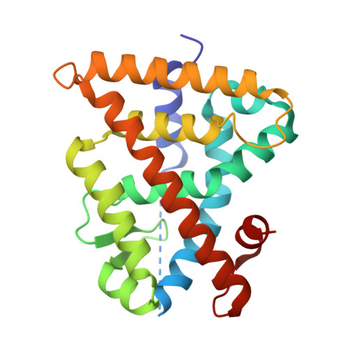
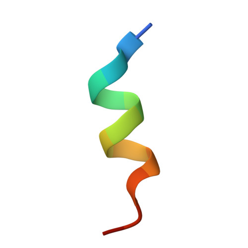
Oxaprozin Analogues as Selective RXR Agonists with Superior Properties and Pharmacokinetics.

Schierle, S.Chaikuad, A.Lillich, F.F.Ni, X.Woltersdorf, S.Schallmayer, E.Renelt, B.Ronchetti, R.Knapp, S.Proschak, E.Merk, D.

(2021) J Med Chem 64: 5123-5136

  • DOI: https://doi.org/10.1021/acs.jmedchem.1c00235
  • Primary Citation of Related Structures:  
    7B88, 7B9O

  • PubMed Abstract: 

    The retinoid X receptors (RXR) are ligand-activated transcription factors involved in multiple regulatory networks as universal heterodimer partners for nuclear receptors. Despite their high therapeutic potential in many pathologies, targeting of RXR has only been exploited in cancer treatment as the currently available RXR agonists suffer from exceptional lipophilicity, poor pharmacokinetics (PK), and adverse effects. Aiming to overcome the limitations and to provide improved RXR ligands, we developed a new potent RXR ligand chemotype based on the nonsteroidal anti-inflammatory drug oxaprozin. Systematic structure-activity relationship analysis enabled structural optimization toward low nanomolar potency similar to the well-established rexinoids. Cocrystal structures of the most active derivatives demonstrated orthosteric binding, and in vivo profiling revealed superior PK properties compared to current RXR agonists. The optimized compounds were highly selective for RXR activation and induced RXR-regulated gene expression in native cellular and in vivo settings suggesting them as excellent chemical tools to further explore the therapeutic potential of RXR.


  • Organizational Affiliation

    Institute of Pharmaceutical Chemistry, Goethe University Frankfurt, Max-von-Laue-Str. 9, D-60438 Frankfurt, Germany.


Macromolecules
Find similar proteins by:  (by identity cutoff)  |  3D Structure
Entity ID: 1
MoleculeChains Sequence LengthOrganismDetailsImage
Retinoic acid receptor RXR-alpha230Homo sapiensMutation(s): 0 
Gene Names: RXRANR2B1
UniProt & NIH Common Fund Data Resources
Find proteins for P19793 (Homo sapiens)
Explore P19793 
Go to UniProtKB:  P19793
PHAROS:  P19793
GTEx:  ENSG00000186350 
Entity Groups  
Sequence Clusters30% Identity50% Identity70% Identity90% Identity95% Identity100% Identity
UniProt GroupP19793
Sequence Annotations
Expand
  • Reference Sequence

Find similar proteins by:  Sequence   |   3D Structure  

Entity ID: 2
MoleculeChains Sequence LengthOrganismDetailsImage
Nuclear receptor coactivator 214Homo sapiensMutation(s): 0 
UniProt & NIH Common Fund Data Resources
Find proteins for Q15596 (Homo sapiens)
Explore Q15596 
Go to UniProtKB:  Q15596
PHAROS:  Q15596
GTEx:  ENSG00000140396 
Entity Groups  
Sequence Clusters30% Identity50% Identity70% Identity90% Identity95% Identity100% Identity
UniProt GroupQ15596
Sequence Annotations
Expand
  • Reference Sequence
Small Molecules
Ligands 1 Unique
IDChains Name / Formula / InChI Key2D Diagram3D Interactions
T2K (Subject of Investigation/LOI)
Query on T2K

Download Ideal Coordinates CCD File 
C [auth A]3-[5-[3,5-bis(chloranyl)phenyl]-4-phenyl-1,3-oxazol-2-yl]propanoic acid
C18 H13 Cl2 N O3
MNBKLGOJIXFSTI-UHFFFAOYSA-N
Experimental Data & Validation

Experimental Data

  • Method: X-RAY DIFFRACTION
  • Resolution: 2.38 Å
  • R-Value Free: 0.259 
  • R-Value Work: 0.199 
  • R-Value Observed: 0.202 
  • Space Group: P 43 21 2
Unit Cell:
Length ( Å )Angle ( ˚ )
a = 64.054α = 90
b = 64.054β = 90
c = 110.067γ = 90
Software Package:
Software NamePurpose
REFMACrefinement
Aimlessdata scaling
PDB_EXTRACTdata extraction
XDSdata reduction
PHASERphasing

Structure Validation

View Full Validation Report


Ligand Structure Quality Assessment 


Entry History 

Deposition Data

Revision History  (Full details and data files)

  • Version 1.0: 2021-01-13
    Type: Initial release
  • Version 1.1: 2021-04-14
    Changes: Database references
  • Version 1.2: 2021-05-05
    Changes: Database references
  • Version 1.3: 2022-08-10
    Changes: Database references, Derived calculations
  • Version 1.4: 2024-01-31
    Changes: Data collection, Refinement description