Super-assembly of ER-phagy receptor Atg40 induces local ER remodeling at contacts with forming autophagosomal membranes.
Mochida, K., Yamasaki, A., Matoba, K., Kirisako, H., Noda, N.N., Nakatogawa, H.(2020) Nat Commun 11: 3306-3306
- PubMed: 32620754 Search on PubMedSearch on PubMed Central
- DOI: https://doi.org/10.1038/s41467-020-17163-y
- Primary Citation Related Structures:
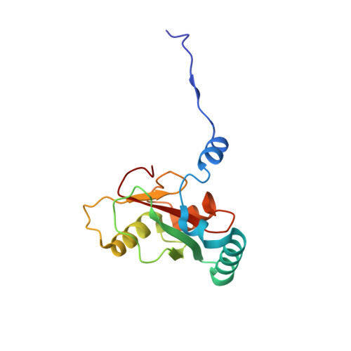
7BRN, 7BRQ, 7BRT, 7BRU - PubMed Abstract:
The endoplasmic reticulum (ER) is selectively degraded by autophagy (ER-phagy) through proteins called ER-phagy receptors. In Saccharomyces cerevisiae, Atg40 acts as an ER-phagy receptor to sequester ER fragments into autophagosomes by binding Atg8 on forming autophagosomal membranes. During ER-phagy, parts of the ER are morphologically rearranged, fragmented, and loaded into autophagosomes, but the mechanism remains poorly understood. Here we find that Atg40 molecules assemble in the ER membrane concurrently with autophagosome formation via multivalent interaction with Atg8. Atg8-mediated super-assembly of Atg40 generates highly-curved ER regions, depending on its reticulon-like domain, and supports packing of these regions into autophagosomes. Moreover, tight binding of Atg40 to Atg8 is achieved by a short helix C-terminal to the Atg8-family interacting motif, and this feature is also observed for mammalian ER-phagy receptors. Thus, this study significantly advances our understanding of the mechanisms of ER-phagy and also provides insights into organelle fragmentation in selective autophagy of other organelles.
- School of Life Science and Technology, Tokyo Institute of Technology, Yokohama, Japan.
Organizational Affiliation: