


Crystal structure of the p53-binding domain of human MdmX protein in complex with Nutlin3a
Cheng, X.Y., Zhang, B.L., Kuang, Z.K., Yang, J., Li, Z.C., Yu, J.P., Zhao, Z.T., Cao, C.Z., Su, Z.D.To be published.
Experimental Data Snapshot
Starting Models: experimental
View more details
Entity ID: 1 | |||||
|---|---|---|---|---|---|
| Molecule | Chains | Sequence Length | Organism | Details | Image |
| Protein Mdm4 | 96 | Homo sapiens | Mutation(s): 0 Gene Names: MDM4, MDMX | 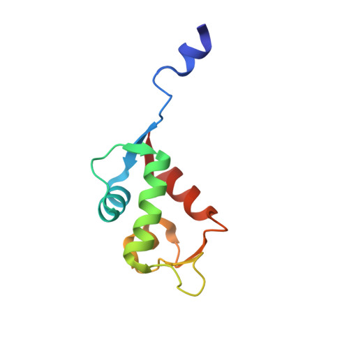 | |
UniProt & NIH Common Fund Data Resources | |||||
PHAROS: O15151 GTEx: ENSG00000198625 | |||||
Entity Groups | |||||
| Sequence Clusters | 30% Identity50% Identity70% Identity90% Identity95% Identity100% Identity | ||||
| UniProt Group | O15151 | ||||
Sequence AnnotationsExpand | |||||
Reference Sequence | |||||
| Ligands 2 Unique | |||||
|---|---|---|---|---|---|
| ID | Chains | Name / Formula / InChI Key | 2D Diagram | 3D Interactions | |
| NUT (Subject of Investigation/LOI) Download:Ideal Coordinates CCD File | B [auth A] | 4-({(4S,5R)-4,5-bis(4-chlorophenyl)-2-[4-methoxy-2-(propan-2-yloxy)phenyl]-4,5-dihydro-1H-imidazol-1-yl}carbonyl)piperazin-2-one C30 H30 Cl2 N4 O4 BDUHCSBCVGXTJM-WUFINQPMSA-N |  | ||
| O4B Download:Ideal Coordinates CCD File | C [auth A] | 1,4,7,10,13,16-HEXAOXACYCLOOCTADECANE C12 H24 O6 XEZNGIUYQVAUSS-UHFFFAOYSA-N |  | ||
| Length ( Å ) | Angle ( ˚ ) |
|---|---|
| a = 47.47 | α = 90 |
| b = 47.47 | β = 90 |
| c = 91.19 | γ = 90 |
| Software Name | Purpose |
|---|---|
| HKL-2000 | data reduction |
| Aimless | data scaling |
| PHASER | phasing |
| REFMAC | refinement |
| PDB_EXTRACT | data extraction |


