7CA5

Cryo-EM structure of human GABA(B) receptor in apo state


Experimental Data Snapshot

  • Method: ELECTRON MICROSCOPY
  • Resolution: 7.60 Å
  • Aggregation State: PARTICLE 
  • Reconstruction Method: SINGLE PARTICLE 

wwPDB Validation   3D Report Full Report


This is version 1.1 of the entry. See complete history


Literature

Structural Basis for Activation of the Heterodimeric GABA B Receptor.

Kim, Y.Jeong, E.Jeong, J.H.Kim, Y.Cho, Y.

(2020) J Mol Biol 432: 5966-5984

  • DOI: https://doi.org/10.1016/j.jmb.2020.09.023
  • Primary Citation of Related Structures:  
    7CA3, 7CA5, 7CUM

  • PubMed Abstract: 

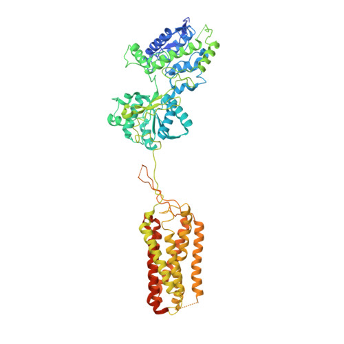
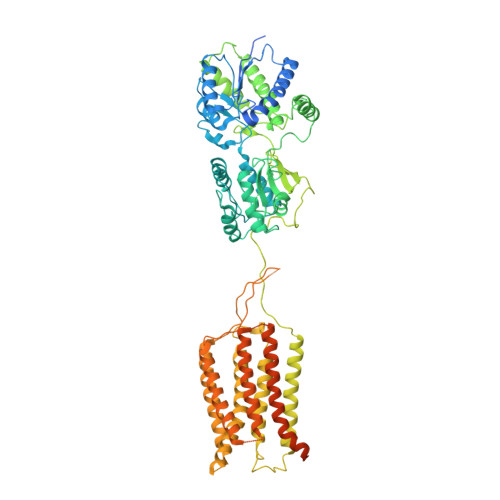
    The neurotransmitter γ-aminobutyric acid (GABA) activates the metabotropic GABA B receptor to generate slow, prolonged inhibitory signals that regulate the neural circuitry. The GABA B receptor is an obligate heterodimeric G protein-coupled receptor (GPCR) comprised of GBR1 and GBR2 subunits, each with extracellular, seven-helix transmembrane (7TM), and coiled-coil domains. To understand how GABA-driven conformational changes in the extracellular domain are transmitted to the 7TM domain during signal transduction, we determined cryo-electron microscopy (EM) structures of GABA B in two different states: an antagonist-bound inactive state, and an active state in which both the GABA agonist and a positive allosteric modulator (PAM) are bound. In the inactive state, the TM3 and TM5 helices in the two 7TM domains engage in cholesterol-mediated as well as direct interactions, resulting in an open conformation. GABA binding forces the extracellular domains of GBR1 and GBR2 into a compact form, relocating the linkers that connect the extracellular and 7TM domains closer to each other. The movement of the linker along with the associated extracellular loop 2 of the 7TM domain reorients the two 7TM domains and creates a new interface with the TM5, TM6 and TM7 helices in a closed conformation. PAM binding to the interface between the TM6 and TM6 helices stabilizes the active 7TM domain conformation. The relayed structural rearrangement results in significant conformational changes in the TM helices, as well as intracellular loop 3 in GBR2, which may promote the binding and activation of the Gi/o proteins.


  • Organizational Affiliation

    Department of Life Science, Pohang University of Science and Technology, Pohang, Republic of Korea.


Macromolecules
Find similar proteins by:  (by identity cutoff)  |  3D Structure
Entity ID: 1
MoleculeChains Sequence LengthOrganismDetailsImage
Gamma-aminobutyric acid type B receptor subunit 1771Homo sapiensMutation(s): 0 
Gene Names: GABBR1GPRC3A
Membrane Entity: Yes 
UniProt & NIH Common Fund Data Resources
Find proteins for Q9UBS5 (Homo sapiens)
Explore Q9UBS5 
Go to UniProtKB:  Q9UBS5
PHAROS:  Q9UBS5
GTEx:  ENSG00000204681 
Entity Groups  
Sequence Clusters30% Identity50% Identity70% Identity90% Identity95% Identity100% Identity
UniProt GroupQ9UBS5
Sequence Annotations
Expand
  • Reference Sequence
Find similar proteins by:  (by identity cutoff)  |  3D Structure
Entity ID: 2
MoleculeChains Sequence LengthOrganismDetailsImage
Gamma-aminobutyric acid type B receptor subunit 2822Homo sapiensMutation(s): 0 
Gene Names: GABBR2GPR51GPRC3B
Membrane Entity: Yes 
UniProt & NIH Common Fund Data Resources
Find proteins for O75899 (Homo sapiens)
Explore O75899 
Go to UniProtKB:  O75899
PHAROS:  O75899
GTEx:  ENSG00000136928 
Entity Groups  
Sequence Clusters30% Identity50% Identity70% Identity90% Identity95% Identity100% Identity
UniProt GroupO75899
Sequence Annotations
Expand
  • Reference Sequence
Experimental Data & Validation

Experimental Data

  • Method: ELECTRON MICROSCOPY
  • Resolution: 7.60 Å
  • Aggregation State: PARTICLE 
  • Reconstruction Method: SINGLE PARTICLE 

Structure Validation

View Full Validation Report


Entry History & Funding Information

Deposition Data


Funding OrganizationLocationGrant Number
Other privateKorea, Republic OfSamsung Science and Technology Foundation SSTF-BA1602-14

Revision History  (Full details and data files)

  • Version 1.0: 2020-11-11
    Type: Initial release
  • Version 1.1: 2024-03-27
    Changes: Data collection, Database references